
-
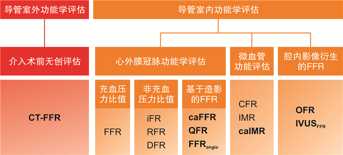
摘要: 冠状动脉功能学评估是经皮冠状动脉介入治疗(PCI)临床决策的主要依据。血流储备分数(FFR)是功能学评估的金标准,但限于操作复杂、耗时长等原因,其临床应用十分有限。随着计算流体力学的发展,基于影像衍生的功能学指标(CT-FFR、QFR、caFFR、caIMR等)应运而生,其无创、高效且诊断准确率较高,在介入术前评估和术中指导中逐渐体现出优势。本文将介绍不同功能学衍生技术的应用场景,并对影像与功能学联合应用的价值进行展望。Abstract: Coronary function assessment has become the cornerstone of clinical decision-making in percutaneous coronary intervention(PCI). Fractional flow reserve(FFR) is the gold standard for functional assessment, but its clinical application is limited due to the complicated and time-consuming procedure. With the development of computational fluid dynamics technology, imaging-derived functional indicators(CT-FFR, QFR, caFFR, caIMR, etc.) have emerged, which are non-invasive, efficient and have high diagnostic accuracy, and have advantages in pre-assessment and guidance of PCI. This article will introduce the application scenarios of different functional derived techniques, and discuss the value of the combined use of imaging and functional techniques.
-
Key words:
- coronary artery disease /
- fractional flow reserve /
- CT-FFR /
- QFR /
- caFFR /
- caIMR
-

-
[1] Pijls NH, van Son JA, Kirkeeide RL, et al. Experimental basis of determining maximum coronary, myocardial, and collateral blood flow by pressure measurements for assessing functional stenosis severity before and after percutaneous transluminal coronary angioplasty[J]. Circulation, 1993, 87(4): 1354-1367. doi: 10.1161/01.CIR.87.4.1354
[2] Pijls NH, van Schaardenburgh P, Manoharan G, et al. Percutaneous coronary intervention of functionally nonsignificant stenosis: 5-year follow-up of the DEFER Study[J]. J Am Coll Cardiol, 2007, 49(21): 2105-2111. doi: 10.1016/j.jacc.2007.01.087
[3] Pijls NH, Fearon WF, Tonino PA, et al. Fractional flow reserve versus angiography for guiding percutaneous coronary intervention in patients with multivessel coronary artery disease: 2-year follow-up of the FAME(Fractional Flow Reserve Versus Angiography for Multivessel Evaluation)study[J]. J Am Coll Cardiol, 2010, 56(3): 177-184. doi: 10.1016/j.jacc.2010.04.012
[4] De Bruyne B, Pijls NH, Kalesan B, et al. Fractional flow reserve-guided PCI versus medical therapy in stable coronary disease[J]. N Engl J Med, 2012, 367(11): 991-1001. doi: 10.1056/NEJMoa1205361
[5] Layland J, Oldroyd KG, Curzen N, et al. Fractional flow reserve vs. angiography in guiding management to optimize outcomes in non-ST-segment elevation myocardial infarction: the British Heart Foundation FAMOUS-NSTEMI randomized trial[J]. Eur Heart J, 2015, 36(2): 100-111. doi: 10.1093/eurheartj/ehu338
[6] Smits PC, Abdel-Wahab M, Neumann FJ, et al. Fractional flow reserve-guided multivessel angioplasty in myocardial infarction[J]. N Engl J Med, 2017, 376(13): 1234-1244. doi: 10.1056/NEJMoa1701067
[7] 韩雅玲. 中国经皮冠状动脉介入治疗指南(2016)[J]. 中华心血管病杂志, 2016, 44(5): 382-400. doi: 10.3760/cma.j.issn.0253-3758.2016.05.006
[8] Neumann FJ, Sousa-Uva M, Ahlsson A, et al. 2018 ESC/EACTS Guidelines on myocardial revascularization[J]. Eur Heart J, 2019, 40(2): 87-165. doi: 10.1093/eurheartj/ehy394
[9] Puymirat E, Cayla G, Simon T, et al. Multivessel PCI Guided by FFR or angiography for myocardial infarction[J]. N Engl J Med, 2021, 385(4): 297-308. doi: 10.1056/NEJMoa2104650
[10] Fearon WF, Zimmermann FM, De Bruyne B, et al. Fractional flow reserve-guided PCI as compared with coronary bypass surgery[J]. N Engl J Med, 2022, 386(2): 128-137. doi: 10.1056/NEJMoa2112299
[11] Kogame N, Ono M, Kawashima H, et al. The impact of coronary physiology on contemporary clinical decision making[J]. JACC Cardiovasc Interv, 2020, 13(14): 1617-1638. doi: 10.1016/j.jcin.2020.04.040
[12] Min JK, Taylor CA, Achenbach S, et al. Noninvasive fractional flow reserve derived from coronary CT angiography: clinical data and scientific principles[J]. JACC Cardiovasc Imaging, 2015, 8(10): 1209-1222. doi: 10.1016/j.jcmg.2015.08.006
[13] Koo BK, Erglis A, Doh JH, et al. Diagnosis of ischemia-causing coronary stenoses by noninvasive fractional flow reserve computed from coronary computed tomographic angiograms. Results from the prospective multicenter DISCOVER-FLOW(Diagnosis of Ischemia-Causing Stenoses Obtained Via Noninvasive Fractional Flow Reserve)study[J]. J Am Coll Cardiol, 2011, 58(19): 1989-1997. doi: 10.1016/j.jacc.2011.06.066
[14] Norgaard BL, Leipsic J, Gaur S, et al. Diagnostic performance of noninvasive fractional flow reserve derived from coronary computed tomography angiography in suspected coronary artery disease: the NXT trial(Analysis of Coronary Blood Flow Using CT Angiography: Next Steps)[J]. J Am Coll Cardiol, 2014, 63(12): 1145-1155. doi: 10.1016/j.jacc.2013.11.043
[15] Driessen RS, Danad I, Stuijfzand WJ, et al. Comparison of coronary computed tomography angiography, fractional flow reserve, and perfusion imaging for ischemia diagnosis[J]. J Am Coll Cardiol, 2019, 73(2): 161-173. doi: 10.1016/j.jacc.2018.10.056
[16] Wang X, Zeng Y, Tang Z, et al. Diagnostic accuracy of computed tomography-based fractional flow reserve with a new coarse-to-fine subpixel algorithm in detecting hemodynamically significant stenosis[P]. Paper presented at: ACC, March 8, 2022, Washington DC.
[17] Douglas PS, Pontone G, Hlatky MA, et al. Clinical outcomes of fractional flow reserve by computed tomographic angiography-guided diagnostic strategies vs. usual care in patients with suspected coronary artery disease: the prospective longitudinal trial of FFR(CT): outcome and resource impacts study[J]. Eur Heart J, 2015, 36(47): 3359-3367. doi: 10.1093/eurheartj/ehv444
[18] Fairbairn TA, Nieman K, Akasaka T, et al. Real-world clinical utility and impact on clinical decision-making of coronary computed tomography angiography-derived fractional flow reserve: lessons from the ADVANCE Registry[J]. Eur Heart J, 2018, 39(41): 3701-3711. doi: 10.1093/eurheartj/ehy530
[19] Hlatky MA, De Bruyne B, Pontone G, et al. Quality-of-life and economic outcomes of assessing fractional flow reserve with computed tomography angiography: PLATFORM[J]. J Am Coll Cardiol, 2015, 66(21): 2315-2323. doi: 10.1016/j.jacc.2015.09.051
[20] Curzen N, Nicholas Z, Stuart B, et al. Fractional flow reserve derived from computed tomography coronary angiography in the assessment and management of stable chest pain: the FORECAST randomized trial[J]. Eur Heart J, 2021, 42(37): 3844-3852. doi: 10.1093/eurheartj/ehab444
[21] Nanna MG, Vemulapalli S, Fordyce CB, et al. The prospective randomized trial of the optimal evaluation of cardiac symptoms and revascularization: Rationale and design of the PRECISE trial[J]. Am Heart J, 2022, 245: 136-148. doi: 10.1016/j.ahj.2021.12.004
[22] Collet C, Onuma Y, Andreini D, et al. Coronary computed tomography angiography for heart team decision-making in multivessel coronary artery disease[J]. Eur Heart J, 2018, 39(41): 3689-3698.
[23] Knuuti J, Wijns W, Saraste A, et al. 2019 ESC Guidelines for the diagnosis and management of chronic coronary syndromes[J]. Eur Heart J, 2020, 41(3): 407-477. doi: 10.1093/eurheartj/ehz425
[24] Xu B, Tu S, Qiao S, et al. Diagnostic accuracy of angiography-based quantitative flow ratio measurements for online assessment of coronary stenosis[J]. J Am Coll Cardiol, 2017, 70(25): 3077-3087. doi: 10.1016/j.jacc.2017.10.035
[25] Li J, Gong Y, Wang W, et al. Accuracy of computational pressure-fluid dynamics applied to coronary angiography to derive fractional flow reserve: FLASH FFR[J]. Cardiovasc Res, 2020, 116(7): 1349-1356. doi: 10.1093/cvr/cvz289
[26] Xu B, Tu S, Song L, et al. Angiographic quantitative flow ratio-guided coronary intervention(FAVOR Ⅲ China): a multicentre, randomised, sham-controlled trial[J]. Lancet, 2021, 398(10317): 2149-2159. doi: 10.1016/S0140-6736(21)02248-0
[27] Li SJ, Ge Z, Kan J, et al. Cutoff value and long-term prediction of clinical events by FFR measured immediately after implantation of a drug-eluting stent in patients with coronary artery disease: 1-to 3-year results from the DKCRUSH Ⅶ Registry Study[J]. JACC Cardiovasc Interv, 2017, 10(10): 986-995. doi: 10.1016/j.jcin.2017.02.012
[28] Biscaglia S, Tebaldi M, Brugaletta S, et al. Prognostic value of QFR measured immediately after successful stent implantation: the international multicenter prospective HAWKEYE Study[J]. JACC Cardiovasc Interv, 2019, 12(20): 2079-2088. doi: 10.1016/j.jcin.2019.06.003
[29] Kogame N, Takahashi K, Tomaniak M, et al. Clinical implication of quantitative flow ratio after percutaneous coronary intervention for 3-vessel disease[J]. JACC Cardiovasc Interv, 2019, 12(20): 2064-2075. doi: 10.1016/j.jcin.2019.08.009
[30] Agarwal SK, Kasula S, Hacioglu Y, et al. Utilizing post-intervention fractional flow reserve to optimize acute results and the relationship to long-term outcomes[J]. JACC Cardiovasc Interv, 2016, 9(10): 1022-1031. doi: 10.1016/j.jcin.2016.01.046
[31] Jeremias A, Davies JE, Maehara A, et al. Blinded physiological assessment of residual ischemia after successful angiographic percutaneous coronary intervention: The DEFINE PCI Study[J]. JACC Cardiovasc Interv, 2019, 12(20): 1991-2001. doi: 10.1016/j.jcin.2019.05.054
[32] Collison D, Didagelos M, Aetesam-Ur-Rahman M, et al. Post-stenting fractional flow reserve vs coronary angiography for optimization of percutaneous coronary intervention(TARGET-FFR)[J]. Eur Heart J, 2021, 42(45): 4656-4668. doi: 10.1093/eurheartj/ehab449
[33] van Zandvoort LJC, Masdjedi K, Tovar Forero MN, et al. Fractional flow reserve guided percutaneous coronary intervention optimization directed by high-definition intravascular ultrasound versus standard of care: Rationale and study design of the prospective randomized FFR-REACT trial[J]. Am Heart J, 2019, 213: 66-72. doi: 10.1016/j.ahj.2019.03.017
[34] Fearon WF, Low AF, Yong AS, et al. Prognostic value of the Index of Microcirculatory Resistance measured after primary percutaneous coronary intervention[J]. Circulation, 2013, 127(24): 2436-2441. doi: 10.1161/CIRCULATIONAHA.112.000298
[35] Nishi T, Murai T, Ciccarelli G, et al. Prognostic value of coronary microvascular function measured immediately after percutaneous coronary intervention in stable coronary artery disease: an international multicenter study[J]. Circ Cardiovasc Interv, 2019, 12(9): e007889. doi: 10.1161/CIRCINTERVENTIONS.119.007889
[36] Ai H, Feng Y, Gong Y, et al. Coronary angiography-derived index of microvascular resistance[J]. Front Physiol, 2020, 11: 605356. doi: 10.3389/fphys.2020.605356
[37] Mejia-Renteria H, Lee JM, Choi KH, et al. Coronary microcirculation assessment using functional angiography: Development of a wire-free method applicable to conventional coronary angiograms[J]. Catheter Cardiovasc Interv, 2021, 98(6): 1027-1037. doi: 10.1002/ccd.29863
[38] Shin D, Kim J, Choi KH, et al. Functional angiography-derived index of microcirculatory resistance validated with microvascular obstruction in cardiac magnetic resonance after STEMI[J]. Rev Esp Cardiol(Engl Ed). 2022.
[39] Choi KH, Dai N, Li Y, et al. Functional coronary angiography-derived index of microcirculatory resistance in patients with ST-segment elevation myocardial infarction[J]. JACC Cardiovasc Interv, 2021, 14(15): 1670-1684. doi: 10.1016/j.jcin.2021.05.027
[40] Tian F, Yu W, Huang J, et al. First presentation of integration of intravascular optical coherence tomography and computational fractional flow reserve[J]. Int J Cardiovasc Imaging, 2019, 35(4): 601-602. doi: 10.1007/s10554-018-1491-1
[41] Yu W, Huang J, Jia D, et al. Diagnostic accuracy of intracoronary optical coherence tomography-derived fractional flow reserve for assessment of coronary stenosis severity[J]. EuroIntervention, 2019, 15(2): 189-197. doi: 10.4244/EIJ-D-19-00182
[42] Gutierrez-Chico JL, Chen Y, Yu W, et al. Diagnostic accuracy and reproducibility of optical flow ratio for functional evaluation of coronary stenosis in a prospective series[J]. Cardiol J, 2020, 27(4): 350-361. doi: 10.5603/CJ.a2020.0071
[43] van Zandvoort LJC, Ali Z, Kern M, et al. Improving PCI outcomes using postprocedural physiology and intravascular imaging[J]. JACC Cardiovasc Interv, 2021, 14(22): 2415-2430. doi: 10.1016/j.jcin.2021.08.069
[44] Seike F, Uetani T, Nishimura K, et al. Intravascular ultrasound-derived virtual fractional flow reserve for the assessment of myocardial ischemia[J]. Circ J, 2018, 82(3): 815-823. doi: 10.1253/circj.CJ-17-1042
[45] Bezerra CG, Hideo-Kajita A, Bulant CA, et al. Coronary fractional flow reserve derived from intravascular ultrasound imaging: Validation of a new computational method of fusion between anatomy and physiology[J]. Catheter Cardiovasc Interv, 2019, 93(2): 266-274. doi: 10.1002/ccd.27822
[46] Yu W, Tanigaki T, Ding D, et al. Accuracy of intravascular ultrasound-based fractional flow reserve in identifying hemodynamic significance of coronary stenosis[J]. Circ Cardiovasc Interv, 2021, 14(2): e009840. doi: 10.1161/CIRCINTERVENTIONS.120.009840
[47] Xaplanteris P, Fournier S, Pijls NHJ, et al. Five-year outcomes with PCI guided by fractional flow reserve[J]. N Engl J Med, 2018, 379(3): 250-259. doi: 10.1056/NEJMoa1803538
[48] Lee JM, Choi KH, Koo BK, et al. Prognostic implications of plaque characteristics and stenosis severity in patients with coronary artery disease[J]. J Am Coll Cardiol, 2019, 73(19): 2413-2424. doi: 10.1016/j.jacc.2019.02.060
[49] Yang S, Koo BK, Narula J. Interactions between morphological plaque characteristics and coronary physiology: from pathophysiological basis to clinical implications[J]. JACC Cardiovasc Imaging, 2021, S1936-878X(21): 00775-0.
-
 下载:
下载:
图(1)
计量
- 文章访问数: 1657
- PDF下载数: 1695
- 施引文献: 0