
A case of descending aorta multiple vegetations diagnosis by three-dimensional transesophageal echocardiography
-
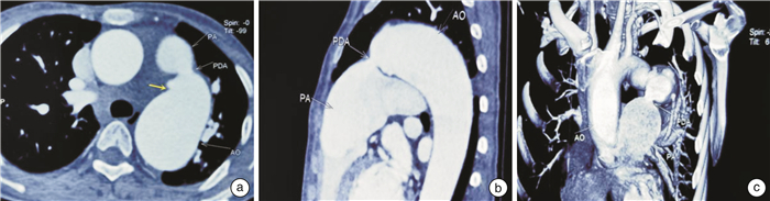
摘要: 本例患者术前经胸超声提示主动脉瓣、动脉导管及降主动脉赘生物形成,对于降主动脉拟行主动弓置换术。通过术前三维经食管超声提示降主动脉赘生物内基本已形成钙化,较为稳定,呈环形分布,未侵及毗邻血管壁,遂未对降主动脉进一步处理,仅行主动脉瓣赘生物清除、主动脉瓣置换、动脉导管缝扎及赘生物清除术。患者术后随访3个月未出现发热、肢体及重要脏器栓塞等症状。
-
关键词:
- 三维经食管超声心动图 /
- 降主动脉 /
- 多发赘生物
Abstract: The patient's preoperative results of transthoracic echocardiography(TTE) indicated that vegetations were formed in the aortic valve, ductus arteriosus and descending aorta. Active arch replacement was planned for treating with the descending aorta. According to preoperative results from three-dimensional transesophageal echocardiography(3D-TEE), calcification was found in the vegetations of the descending aorta which was relatively stable. The calcification was distributed in a "ring" shape without adjacent blood vessels invaded. Therefore, no further treatment was performed on the descending aorta while aortic valve vegetation dissection, aortic valve replacement, ligation of patent ductus arteriosus and dissection of vegetations were performed. The followed-up was conducted for three months after the operation which no symptoms such as fever, limb and important organ embolism. -

-
[1] Rajani R, Klein JL. Infective endocarditis: A contemporary update[J]. Clin Med(Lond), 2020, 20(1): 31-35.
[2] 中华医学会心血管病学分会. 成人感染性心内膜炎预防、诊断和治疗专家共识[J]. 中华心血管病杂志, 2014, 42(10): 806-816. doi: 10.3760/cma.j.issn.0253-3758.2014.10.004
[3] Yuan XC, Liu M, Hu J, et al. Diagnosis of infective endocarditis using echocardiography[J]. Medicine(Baltimore), 2019, 98(38): e17141.
[4] Gnanam D, Bartelds B, van Leeuwen WJ, et al. A case report on endarteritis in a child with coarctation of aorta[J]. Echocardiography, 2019, 36(7): 1427-1430. doi: 10.1111/echo.14418
[5] 张海波, 李守军. 先天性心脏病外科治疗中国专家共识(十一): 主动脉缩窄与主动脉弓中断[J]. 中国胸心血管外科临床杂志, 2020, 27(11): 1255-1261. https://www.cnki.com.cn/Article/CJFDTOTAL-ZXYX202011003.htm
[6] 吴正华, 周爱云, 张诚, 等. 经食管超声心动图监测动脉导管未闭封堵术的应用价值[J]. 临床心血管病杂志, 2014, 30(12): 1030-1032. https://www.cnki.com.cn/Article/CJFDTOTAL-LCXB201412004.htm
[7] 洪灿, 刘素君, 周佳, 等. 经食管三维超声心动图对感染性心内膜炎赘生物的诊断价值[J]. 中华医院感染学杂志, 2017, 27(17): 3867-3870. https://www.cnki.com.cn/Article/CJFDTOTAL-ZHYY201717009.htm
[8] Baddour LM, Wilson WR, Bayer AS, et al. Infective endocarditis in adults: diagnosis, antimicrobial therapy, and management of complications: a scientific statement for healthcare professionals from the american heart association[J]. Circulation, 2015, 132(15): 1435-1486. doi: 10.1161/CIR.0000000000000296
[9] Kim IC, Chang S, Hong GR, et al. Comparison of cardiac computed tomography with transesophageal echocardiography for identifying vegetation and intracardiac complications in patients with infective endocarditis in the era of 3-dimensional images[J]. Circ Cardiovasc Imaging, 2018, 11(3): e006986.
[10] Gomes A, van Geel PP, Santing M, et al. Imaging infective endocarditis: Adherence to a diagnostic flowchart and direct comparison of imaging techniques[J]. J Nucl Cardiol, 2020, 27(2): 592-608.
[11] 中华医学会胸心血管外科分会瓣膜病外科学组. 感染性心内膜炎外科治疗中国专家共识[J]. 中华胸心血管外科杂志, 2022, 38(3): 146-155. https://www.cnki.com.cn/Article/CJFDTOTAL-LCXB202209003.htm
[12] Al-Riyami AZ, Baskaran B, Panchatcharam SM, et al. Preoperative Anemia is Associated with Increased Intraoperative Mortality in Patients Undergoing Cardiac Surgery[J]. Oman Med, 2021, 36(3): e267.
[13] 李昱茜, 孟欣, 白炜, 等. 三维食管超声指导下经导管主动脉瓣置换术治疗感染性心内膜炎致主动脉瓣关闭不全1例[J]. 临床心血管病杂志, 2022, 38(7): 592-595. https://www.cnki.com.cn/Article/CJFDTOTAL-LCXB202207015.htm
-
 下载:
下载:


图(5)
计量
- 文章访问数: 1155
- PDF下载数: 212
- 施引文献: 0