
-
摘要: 目的 分析多模态影像学技术在儿童冠状动脉(冠脉)起源异常中的诊断价值。方法 回顾分析我院近5年确诊为冠脉起源异常患儿的经胸超声心动图(TTE)、冠脉CTA、心脏增强磁共振(MRI)、冠脉造影(CA)等检查结果、图像及病历资料。结果 冠脉起源异常患儿63例,其中冠脉异常起源于肺动脉(ACAPA)26例,年龄1个月~10岁,中位年龄6个月;冠脉异常主动脉起源(AAOCA)37例,年龄2个月~15岁,中位年龄7岁。TTE对ACAPA首诊检出率100%,确诊率77%(20/26);对AAOCA首诊检出率65%(24/37),确诊率46%(17/37),漏诊率35%(13/37)。TTE对心腔内瓣膜等结构、血流动力学和心功能评估有优势。CTA可明确显示冠脉起源位置,缺陷在于对心腔内结构的显示及血流动力学的评估方面,并有放射性。MRI的优势在于评估心肌缺血及是否发生纤维化。CA的优势在于明确冠脉起源位置以及血流充盈情况,缺点是有创和放射性。结论 在儿童冠脉起源异常的诊断中,各项影像学技术各有优势。TTE可作为筛查和首诊的首选技术。Abstract: Objective To analyze the diagnostic value of multimodal imaging technology in children with abnormal coronary artery origin.Methods The results, images of transthoracic echocardiography(TTE), coronary CTA, cardiac contrast magnetic resonance imaging(MRI), coronary angiography(CA) and medical records of children diagnosed with coronary artery abnormalities in our hospital in the past 5 years were retrospectively analyzed.Results There were 63 children with abnormal coronary artery origin, including 26 cases with abnormal coronary artery origin of pulmonary artery(ACAPA), aged 1 month-10 years old, and median age was 6 months; 37 cases of anomalous aortic origin of coronary artery(AAOCA), aged 2 months to 15 years, and median age was 7 years. The initial detection rate of ACAPA by TTE was 100% and the diagnosis rate was 77%(20/26). The initial detection rate of AAOCA was 65%(24/37), the diagnosis rate was 46%(17/37), and the missed diagnosis rate was 35%(13/37). TTE has advantages for assessing structures such as valves in the heart chamber, hemodynamics, and cardiac function. CTA can clearly show the location of coronary origin, but it is defective in the visualization of intra-chamber structures and hemodynamic assessment, and has radioactivity. MRI has the advantage of assessing myocardial ischemia and the presence of fibrosis. AC has the advantage of the location of the origin of the coronary artery and the filling of blood flow, but the disadvantage is invasive and radioactive.Conclusion In the diagnosis of abnormal origin of coronary arteries in children, various imaging techniques have their own advantages. TTE is the preferred examination method for screening and initial diagnosis.
-
Key words:
- children /
- abnormal coronary artery origin /
- multimodal imaging /
- echocardiography
-

-
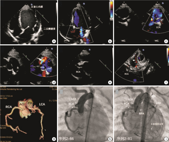
图 1 ALCAPA患儿超声心动图、CTA和CA
Figure 1. Echocardiography, CTA, and CA in children with ALCAPA
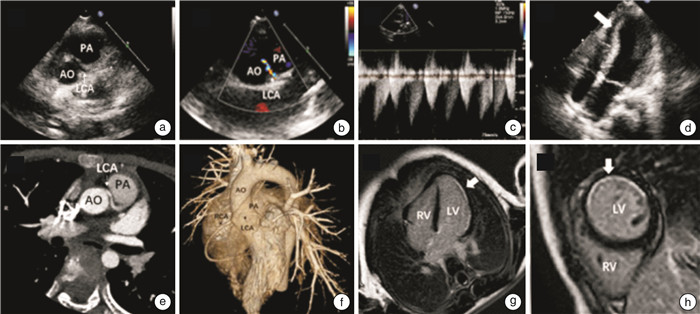
图 2 左冠脉异常起源于右冠窦的超声心动图、CTA、MRI图像
Figure 2. Echocardiography, CTA, and MRI images of left coronary artery abnormalities originating from the right coronary sinus
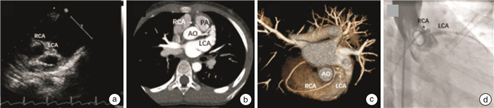
图 3 右冠脉异常起源于左冠窦患儿超声心动图、CTA及冠脉造影图像
Figure 3. Echocardiography, CTA and coronary angiography in children with right coronary artery abnormalities originated from left coronary sinus
表 1 不同影像学技术对本组冠脉起源异常患儿的应用和诊断
Table 1. Application and diagnosis of different imaging techniques in abnormal coronary origin
例(%) 病种 总例数 CTA TTE CA MRI 确诊 疑似 漏诊 ALCAPA 24 23(95.8) 18(75.0) 6(25.0) 0 1(4.2) 0 ARCAPA 2 2(100.0) 2(100.0) 0 0 0 0 ALCA 5 5(100.0) 3(60.0) 0 2(40.0) 0 1(20.0) ARCA 32 32(100.0) 14(43.8) 7(21.9) 11(34.4) 2(6.3) 0  下载: 导出CSV
表 2 ALCAPA患儿TTE表现
Table 2. TTE in ALCAPA children
超声征像 例(%) 直接征象 肺动脉壁探及左冠开口 24(100) 左冠血流逆灌入肺动脉 24(100) 间接征象 左室内径增大 无-轻度 4(16.7) 中-重度 20(83.3) 二尖瓣、腱索、乳头肌 回声增强 24(100) 二尖瓣关闭不全 轻度 7(29.2) 中-重度 17(70.8) LVEF ≥55% 5(20.8) 30%~55% 13(54.2) ≤30% 6(25.0) 心肌内侧支血管 少 8(33.3) 多 16(66.7) 节段性室壁运动异常 3(12.5) 右冠增宽 18(75.0)
下载: 导出CSV
-
[1] Hoffman JI, Kaplan S, Liberthson RR. Prevalence of congenital heart disease[J]. Am Heart J, 2004, 147(3): 425-439. doi: 10.1016/j.ahj.2003.05.003
[2] Castelvecchio S, Pappalardo OA, Menicanti L. Myocardial reconstruction in ischaemic cardiomyopathy[J]. Eur J Cardiothorac Surg, 2019, 55(Suppl 1): i49-i56.
[3] Patel SG, Frommelt MA, Frommelt PC, et al. Echocardiographic diagnosis, surgical treatment, and outcomes of anomalous left coronary artery from the pulmonary artery[J]. J Am Soc Echocardiogr, 2017, 30(9): 896-903. doi: 10.1016/j.echo.2017.05.005
[4] Wesselhoeft H, Fawcett JS, Johnson AL. Anomalous origin of the left coronary artery from the pulmonary trunk. Its clinical spectrum, pathology, and pathophysiology, based on a review of 140 cases with seven further cases[J]. Circulation, 1968, 38(2): 403-425. doi: 10.1161/01.CIR.38.2.403
[5] Kanoh M, Inai K, Shinohara T, et al. Outcomes from anomalous origin of the left coronary artery from the pulmonary artery repair: Long-term complications in relation to residual myocardial abnormalities[J]. J Cardiol, 2017, 70(5): 498-503. doi: 10.1016/j.jjcc.2017.03.008
[6] Cheezum MK, Liberthson RR, Shah NR, et al. Anomalous aortic origin of a coronary artery from the inappropriate sinus of valsalva[J]. J Am Coll Cardiol, 2017, 69(12): 1592-1608. doi: 10.1016/j.jacc.2017.01.031
[7] Brothers JA. Introduction to anomalous aortic origin of a coronary artery[J]. Congenit Heart Dis, 2017, 12(5): 600-602. doi: 10.1111/chd.12497
[8] 安琪, 李守军. 先天性心脏病外科治疗中国专家共识(十二): 先天性冠状动脉异常[J]. 中国胸心血管外科临床杂志, 2020, 12: 1375-1381. https://www.cnki.com.cn/Article/CJFDTOTAL-ZXYX202012002.htm
[9] Herrmann JL, Goldberg LA, Khan AM, et al. A Comparison of perioperative management of anomalous aortic origin of a coronary artery between an adult and pediatric cardiac center[J]. World J Pediatr Congenit Heart Surg, 2016, 7(6): 721-726. doi: 10.1177/2150135116668333
[10] Jegatheeswaran A, Alsoufi B. Anomalous aortic origin of a coronary artery: 2020 year in review[J]. J Thorac Cardiovasc Surg, 2021, 162(2): 353-359. doi: 10.1016/j.jtcvs.2021.04.048
[11] 康彧, 唐红, 赁可. 超声心动图诊断左冠状动脉异常起源于肺动脉2例[J]. 中华医学超声杂志(电子版), 2007, 7(2): 286-288. https://www.cnki.com.cn/Article/CJFDTOTAL-ZHCD201002021.htm
[12] Simon L, Santosh C, Irene DL, et al. Utility of multimodality imaging in the morphologic characterization of anomalous aortic origin of a coronary artery[J]. World J Ped Congen Heart Surg, 2016, 7(3): 308-317. doi: 10.1177/2150135116634326
[13] Yuko O, Nobuko K, Yuzo Y, et al. Evaluation of coronary artery variations using dual-source coronary computed tomography angiography in neonates with transposition of the great arteries[J]. Japan J Radiol, 2019, 37(4): 308-314. doi: 10.1007/s11604-018-00807-x
[14] Goo HW. Identification of coronary artery anatomy on dual-source cardiac computed tomography before arterial switch operation in newborns and young infants: comparison with transthoracic echocardiography[J]. Pediatr Radiol, 2018, 48(2): 176-185. doi: 10.1007/s00247-017-4004-9
[15] Tangcharoen T, Bell A, Hegde S, et al. Detection of coronary artery anomalies in infants and young children with congenital heart disease by using MR imaging[J]. Radiology, 2011, 259(1): 240-247. doi: 10.1148/radiol.10100828
[16] Juan CC, Hwang B, Lee PC, et al. Diagnostic application of multidetector-row computed tomographic coronary angiography to assess coronary abnormalities in pediatric patients: comparison with invasive coronary angiography[J]. Pediatr Neonatol, 2011, 52(4): 208-213. doi: 10.1016/j.pedneo.2011.05.011
-
图(3)
表(2)
计量
- 文章访问数: 805
- PDF下载数: 229
- 施引文献: 0