
-
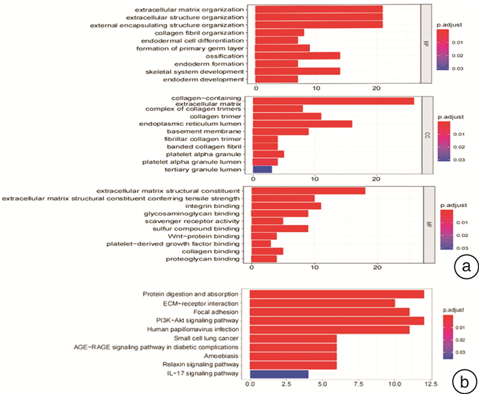
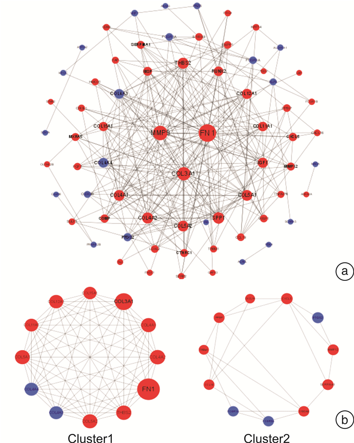
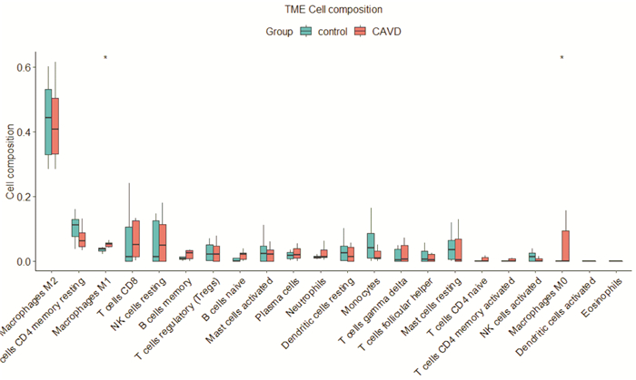
摘要: 目的 探索可能引起钙化性主动脉瓣疾病(CAVD)的关键基因和信号转导通路,寻找CAVD患者瓣膜组织中浸润的关键细胞,为CAVD诊疗提供新的思路。方法 从GEO数据库中获取人类CAVD现有信息芯片,筛选出正常主动脉瓣与CAVD瓣膜之间的差异表达基因(DEGs)。对DEGs的功能和通路基于基因本体论(GO)、京都基因和基因组百科全书(KEGG)进行富集分析。应用String数据库和Cytoscape软件构建蛋白质-蛋白质相互作用(PPI)网络,并筛选出关键基因。运用CIBERSORT数据包计算CAVD中22种免疫细胞浸润比例。结果 通过文本挖掘和数据分析,CAVD患者瓣膜组织有95个DEGs,其中64个基因上调,31个基因下调。这些DEGs通过GO分析富集到细胞外基质组织、细胞外结构组织、外部封装结构组织等117个生物学过程,含胶原蛋白的细胞外基质、内质网腔、胶原蛋白三聚体等13个细胞组分和细胞外基质结构成分、整合素结合、具有拉伸强度的细胞外基质结构成分等33个分子功能。KEGG分析显示主要在蛋白质消化与吸收、细胞外基质-受体相互作用、局灶性黏附等10个方面富集。通过PPI分析筛选出10个关键基因(COL4A2、COL4A1、COL3A1、THBS2、COL5A1、COL5A2、COL4A4、COL11A1、FN1和COL4A3)和两个基因模块,这些关键基因主要在细胞外基质-受体相互作用、黏着斑和蛋白质消化与吸收等方面富集。经过CIBERSORT分析表明CAVD患者M0型巨噬细胞和M1型巨噬细胞细胞水平较高(P < 0.05)。结论 本研究发现FN1、MMP9、COL3A1、SPP1、COL4A2、THBS2、COL5A2、COL5A1、COL4A1和IGF1是CAVD进展中的关键基因。CAVD的发生发展与细胞外基质重塑、巨噬细胞浸润密切相关。细胞外基质调控基因可能可以作为早期CAVD的生物标志物以及CAVD防治的靶标。Abstract: Objective This study in order to explore the key genes and signal transduction pathways that may cause calcific aortic valve disease(CAVD), find the immune cells that infiltrate the valve tissue of CAVD patients, thus, provide new ideas for the provention and treatment of CAVD.Methods The existing information chips of human CAVD were obtained from GEO database to screen the differentially expressed genes(DEGs) between normal aortic valve and calcific aortic valve. The function and pathway of DEGs were analyzed based on gene Ontology(GO) and Kyoto Encyclopedia of Genes and Genomes(KEGG). Use String database and Cytoscape construct Protein-protein interaction(PPI) network, and screen the hub genes. CIBERSORT data package was used to calculate the proportion of 22 kinds of immune cell infiltration in CAVD.Results Through text mining and data analysis, 95 DEGs were found in valve tissues of patients with CAVD, of which 64 genes were up-regulated and 31 genes were down-regulated. These DEGs were enriched into 117 biological processes such as extracellular matrix, extracellular structure and external encapsulation structure through GO analysis. There are 13 components of extracellular matrix containing collagen, endoplasmic reticulum lumen, collagen trimer and 33 molecular functions of extracellular matrix including structural components of extracellular matrix, integrin binding and tensile strength structural components of extracellular matrix. KEGG analysis showed that protein digestion and absorption, extracellular matrix-receptor interaction, focal adhesion and other 10 aspects of enrichment. Ten hub genes(COL4A2, COL4A1, COL3A1, THBS2, COL5A1, COL5A2, COL4A4, COL11A1, FN1 and COL4A3) and two gene modules were screened by PPI analysis. These hub genes are mainly enriched in extracellular matrix-receptor interaction, adhesion plaque and protein digestion and absorption. CIBERSORT analysis showed that the levels of M0 and M1 macrophages were higher in CAVD patients(P < 0.05).Conclusion In this study, FN1, MMP9, COL3A1, SPP1, COL4A2, THBS2, COL5A2, COL5A1, COL4A1 and IGF1 were found as hub genes in the progression of CAVD. The occurrence and development of CAVD are closely related to extracellular matrix remodeling and macrophage infiltration. Extracellular matrix regulation genes may be used as biomarkers for early CAVD and targets for prevention and treatment of CAVD.
-

-
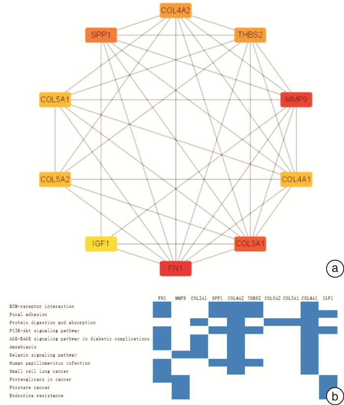
图 4 关键基因的PPI网络和KEGG富集途径
Figure 4. PPI network and KEGG enrichment pathway of key genes
-
[1] Yadgir S, Johnson CO, Aboyans V, et al. Global, regional, and national burden of calcific aortic valve and degenerative mitral valve diseases, 1990-2017[J]. Circulation, 2020, 141(21): 1670-1680. doi: 10.1161/CIRCULATIONAHA.119.043391
[2] Yi B, Zeng W, Lv L, et al. Changing epidemiology of calcific aortic valve disease: 30-year trends of incidence, prevalence, and deaths across 204 countries and territories[J]. Aging(Albany NY), 2021, 13(9): 12710-12732.
[3] 林锐, 王媛, 周宁, 等. 机器学习确定特征性生物标志物预测主动脉瓣置换术后不良心血管事件[J]. 临床心血管病杂志, 2021, 37(3): 248-253. https://www.cnki.com.cn/Article/CJFDTOTAL-LCXB202103013.htm
[4] Blaser MC, Kraler S, Lüscher TF, et al. Multi-omics approaches to define calcific aortic valve disease pathogenesis[J]. Circ Res, 2021, 128(9): 1371-1397. doi: 10.1161/CIRCRESAHA.120.317979
[5] Kostyunin AE, Yuzhalin AE, Ovcharenko EA, et al. Development of calcific aortic valve disease: Do we know enough for new clinical trials?[J]. J Mol Cell Cardiol, 2019, 132: 189-209. doi: 10.1016/j.yjmcc.2019.05.016
[6] Youssef A, Clark JR, Koschinsky ML, et al. Lipoprotein(a): Expanding our knowledge of aortic valve narrowing[J]. Trends Cardiovasc Med, 2021, 31(5): 305-311. doi: 10.1016/j.tcm.2020.06.001
[7] Shuvy M, Abedat S, Eliaz R, et al. Hyperphosphatemia is required for initiation but not propagation of kidney failure-induced calcific aortic valve disease[J]. Am J Physiol Heart Circ Physiol, 2019, 317(4): H695-H704. doi: 10.1152/ajpheart.00765.2018
[8] Edgar R, Domrachev M, Lash AE. Gene expression omnibus: NCBI gene expression and hybridization array data repository[J]. Nucleic Acids Res, 2002, 30(1): 207-210. doi: 10.1093/nar/30.1.207
[9] Kostyunin A, Mukhamadiyarov R, Glushkova T, et al. Ultrastructural pathology of atherosclerosis, calcific aortic valve disease, and bioprosthetic heart valve degeneration: commonalities and differences[J]. Int J Mol Sci, 2020, 21(20): 100.
[10] Boyle EA, Li YI, Pritchard JK. An expanded view of complex traits: from polygenic to omnigenic[J]. Cell, 2017, 169(7): 1177-1186. doi: 10.1016/j.cell.2017.05.038
[11] Kirkness MW, Lehmann K, Forde NR. Mechanics and structural stability of the collagen triple helix[J]. Curr Opin Chem Biol, 2019, 53: 98-105. doi: 10.1016/j.cbpa.2019.08.001
[12] Uchida Y, Shimoyama E, Hiruta N, et al. Detection of early stage of human coronary atherosclerosis by angioscopic imaging of collagen subtypes[J]. J Cardiol, 2021, 77(5): 452-456. doi: 10.1016/j.jjcc.2020.09.011
[13] Hutson HN, Marohl T, Anderson M, et al. Calcific aortic valve disease is associated with layer-specific alterations in collagen architecture[J]. PLoS One, 2016, 11(9): e0163858. doi: 10.1371/journal.pone.0163858
[14] Perrotta I, Davoli M. Collagen mineralization in human aortic valve stenosis: a field emission scanning electron microscopy and energy dispersive spectroscopy analysis[J]. Ultrastruct Pathol, 2014, 38(4): 281-284. doi: 10.3109/01913123.2014.901468
[15] 李春芝, 闫莉, 赵茜, 等. 老年心脏瓣膜钙化与血清胰岛素样生长因子-1的相关性[J]. 临床心血管病杂志, 2015, 31(4): 425-427. https://www.cnki.com.cn/Article/CJFDTOTAL-LCXB201504022.htm
[16] Xiao H, Huang X, Wang S, et al. Metformin ameliorates bleomycin-induced pulmonary fibrosis in mice by suppressing IGF-1[J]. Am J Transl Res, 2020, 12(3): 940-949. doi: 10.1097/01.ccm.0000643508.83802.5f
[17] Swaminathan G, Krishnamurthy VK, Sridhar S, et al. Hypoxia stimulates synthesis of neutrophil gelatinase-associated lipocalin in aortic valve disease[J]. Front Cardiovasc Med, 2019, 6: 156. doi: 10.3389/fcvm.2019.00156
[18] Perrotta I, Sciangula A, Aquila S, et al. Matrix metalloproteinase-9 expression in calcified human aortic valves: a histopathologic, immunohistochemical, and ultrastructural study[J]. Appl Immunohistochem Mol Morphol, 2016, 24(2): 128-137. doi: 10.1097/PAI.0000000000000144
[19] Zhou P, Li Q, Su S, et al. Interleukin 37 suppresses M1 macrophage Polarization through inhibition of the Notch1 and Nuclear Factor Kappa B pathways[J]. Front Cell Dev Biol, 2020, 8: 56. doi: 10.3389/fcell.2020.00056
[20] Chen Y, Waqar AB, Nishijima K, et al. Macrophage-derived MMP-9 enhances the progression of atherosclerotic lesions and vascular calcification in transgenic rabbits[J]. J Cell Mol Med, 2020, 24(7): 4261-4274. doi: 10.1111/jcmm.15087
[21] Grim JC, Aguado BA, Vogt BJ, et al. Secreted factors from proinflammatory macrophages promote an osteoblast-like phenotype in valvular interstitial cells[J]. Arterioscler Thromb Vasc Biol, 2020, 40(11): e296-e308. doi: 10.1161/ATVBAHA.120.315261
[22] Yu X, Zhang QQ, Wang B, et al. Expression and significance of integrin α5β1 and fibronectin in atherosclerotic plaques from autopsy specimens[J]. Zhonghua Bing Li Xue Za Zhi, 2017, 46(3): 182-186. doi: 10.3760/cma.j.issn.0529-5807.2017.03.008
[23] Valiente-Alandi I, Potter SJ, Salvador AM, et al. Inhibiting fibronectin attenuates fibrosis and improves cardiac function in a model of heart failure[J]. Circulation, 2018, 138(12): 1236-1252.
[24] Grau JB, Poggio P, Sainger R, et al. Analysis of osteopontin levels for the identification of asymptomatic patients with calcific aortic valve disease[J]. Ann Thorac Surg, 2012, 93(1): 79-86. doi: 10.1016/j.athoracsur.2011.08.036
[25] Lutz M, von Ingersleben N, Lambers M, et al. Osteopontin predicts clinical outcome in patients after treatment of severe aortic stenosis with transcatheter aortic valve implantation(TAVI)[J]. Open Heart, 2017, 4(2): e000633. doi: 10.1136/openhrt-2017-000633
[26] Li J, Yousefi K, Ding W, et al. Osteopontin RNA aptamer can prevent and reverse pressure overload-induced heart failure[J]. Cardiovasc Res, 2017, 113(6): 633-643. doi: 10.1093/cvr/cvx016
[27] Hsu CH, Liu IF, Kuo HF, et al. miR-29a-3p/THBS2 axis regulates PAH-induced cardiac fibrosis[J]. Int J Mol Sci, 2021, 22(19): 100.
-
 下载:
下载:
图(5)
计量
- 文章访问数: 2663
- PDF下载数: 2423
- 施引文献: 0