
-
摘要: 目的 探究冠心病患者经皮冠状动脉介入治疗(PCI)后支架内再狭窄(ISR)的危险因素,以及磷脂和鞘磷脂代谢差异与ISR的关系。方法 纳入2017年1月—2020年12月在北京安贞医院进行首次PCI手术的冠心病患者共412例,根据PCI术后1年是否发生ISR分为ISR组(35例)和非ISR组(377例)。运用液相色谱串联质谱(LC-MS/MS)技术分析比对两组患者血液样本的代谢产物差异。采用多因素logistic回归分析筛选冠心病患者PCI后ISR的危险因素。结果 非ISR组与ISR组在高密度脂蛋白胆固醇(HDL-C)、血管病变数量、病变长度、支架内径、支架长度、后扩直径方面均差异有统计学意义(均P < 0.05)。多因素logistic回归分析及代谢组学分析结果显示,非ISR组与ISR组患者血液代谢产物存在显著差异,主要为磷脂及鞘磷脂类代谢物的差异(均P < 0.05)。结论 HDL-C、病变长度、支架内径及支架长度为冠心病患者PCI后ISR的影响因素,同时磷脂及鞘磷脂类代谢物在预测ISR的发生方面可能发挥着重要作用。Abstract: Objective To investigate the risk factors for in-stent restenosis(ISR) after percutaneous coronary intervention(PCI) in patients with coronary heart disease, and the relationship between differences in phospholipid and sphingolipid metabolism with ISR.Methods A total of 412 patients with coronary heart disease who underwent their first PCI at Beijing Anzhen Hospital from January 2017 to December 2020 were included, and all patients were divided into the ISR group(n=35) and the non-ISR group(n=377) according to whether ISR occurred within 1 year after PCI. The differences in metabolites of patients' blood samples between the two groups were analyzed using liquid chromatography tandem mass spectrometry(LC-MS/MS). Multi-factor logistic regression was used to analyze the risk factors for ISR after PCI in patients with coronary heart disease.Results There were significant differences in high-density lipoprotein cholesterol(HDL-C), number of vascular lesions, lesion length, stent inner diameter, stent length, and post-dilation diameter between the ISR group and the non-ISR group(all P < 0.05). Multi-factor logistic regression analysis and metabolomic analysis showed that there were significant differences in blood metabolites between the two groups, mainly the differences in phospholipids and sphingomyelin metabolites (all P < 0.05).Conclusion HDL-C, lesion length, stent inner diameter, and stent length are influencing factors of ISR after PCI in patients with coronary heart disease, and phospholipids and sphingolipid metabolites may play an important role in predicting the occurrence of ISR.
-
Key words:
- percutaneous coronary intervention /
- in-stent restenosis /
- metabolomics /
- phospholipids /
- sphingomyelin
-

-
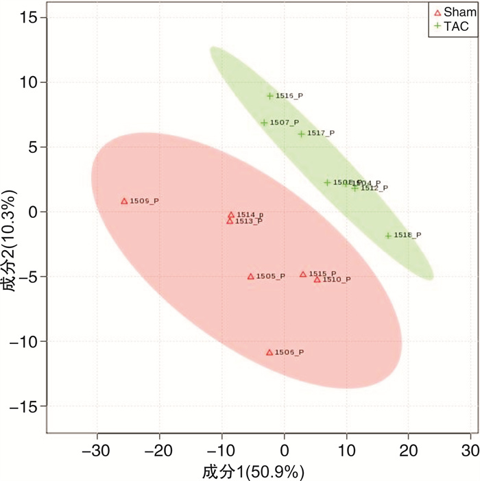
图 1 ISR组和非ISR组血液代谢产物的PLS-DA分析结果
Figure 1. Blood metabolites in non-ISR group and ISR group showed by PLS-DA analysis
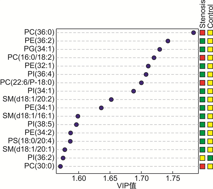
图 2 VIP分析显示的ISR组和非ISR组具有显著差异的代谢产物
Figure 2. Significantly different metabolites between non-ISR group and ISR group showed by VIP analysis
表 1 非ISR组与ISR组基础资料比较
Table 1. Comparison of baseline data between non-ISR group and ISR group
例(%), X±S 项目 非ISR组(377例) ISR组(35例) P 男/女/例 287/90 24/11 0.320 年龄/岁 57.46±9.38 59.29±7.48 0.265 体重指数/(kg·m-2) 25.84±3.28 26.59±3.46 0.197 高血压 121(32.10) 7(20.00) 0.139 糖尿病 117(31.03) 13(37.14) 0.457 吸烟史 205(54.38) 18(51.43) 0.738 饮酒史 128(33.95) 14(40.00) 0.471 HDL-C/(mmol·L-1) 1.26±0.25 1.17±0.28 0.035 LDL-C/(mmol·L-1) 2.48±0.66 2.65±0.58 0.128 TG/(mmol·L-1) 1.22±0.71 1.40±0.67 0.147 hs-CRP/(mg·L-1) 1.11±1.19 1.35±0.96 0.256 CREA/(μmol·L-1) 70.81±15.13 67.39±14.06 0.200 HbA1c/% 6.40±1.21 6.63±1.54 0.313 WBC/(×109·L-1) 6.96±1.89 6.98±1.56 0.967 RBC/(×1012·L-1) 4.76±0.46 4.66±0.48 0.212 病变情况 0.024 单支病变 98(25.99) 5(14.29) 双支病变 141(37.40) 9(25.71) 多支病变 138(36.60) 21(60.00) 病变长度/mm 12.00±5.30 14.81±6.56 0.003 支架数目/个 1.40±0.69 1.49±0.61 0.478 支架内径/mm 2.99±0.46 2.75±0.35 0.002 支架长度/mm 15.38±8.32 18.51±8.20 0.034 预扩直径/mm 2.54±0.41 2.43±0.31 0.127 后扩直径/mm 3.19±0.49 3.02±0.43 0.045  下载: 导出CSV
表 2 ISR组和非ISR组差异性代谢产物
Table 2. Differential metabolites in ISR and non-ISR patients
差异代谢物 VIP P值 变化趋势 PC(36:0) 1.789 0.016 升高 PE(36:2) 1.742 0.021 下降 PG(34:1) 1.729 0.047 下降 PC(16:0/18:2) 1.720 0.027 升高 PE(32:1) 1.711 0.028 下降 PI(36:4) 1.707 0.032 下降 PI(34:1) 1.674 0.019 下降 SM(d18:1/20:2) 1.653 0.025 下降
下载: 导出CSV
表 3 ISR危险因素的logistic分析
Table 3. Logistic analysis of risk factors for ISR
危险因素 OR 95%CI P PC(36:0) 2.94 2.41~3.22 < 0.001 PC(16:0/18:2) 1.28 1.17~1.65 0.010 PE(36:2) 0.42 0.38~0.59 < 0.001 PI(36:4) 0.51 0.34~0.68 0.017 SM(d18:1/20:2) 0.72 0.58~0.82 0.038
下载: 导出CSV
-
[1] Niccoli G, Montone RA, Lanza GA, et al. Angina after percutaneous coronary intervention: The need for precision medicine[J]. Int J Cardiol, 2017, 248: 14-19. doi: 10.1016/j.ijcard.2017.07.105
[2] Li M, Hou J, Gu X, et al. Incidence and risk factors of in-stent restenosis after percutaneous coronary intervention in patients from southern China[J]. Eur J Med Res, 2022, 27(1): 12. doi: 10.1186/s40001-022-00640-z
[3] Moussa ID, Mohananey D, Saucedo J, et al. Trends and outcomes of restenosis after coronary stent implantation in the united states[J]. J Am Coll Cardiol, 2020, 76(13): 1521-1531. doi: 10.1016/j.jacc.2020.08.002
[4] Giacoppo D, Alfonso F, Xu B, et al. Drug-coated balloon angioplasty versus drug-eluting stent implantation in patients with coronary stent restenosis[J]. J Am Coll Cardiol, 2020, 75(21): 2664-2678. doi: 10.1016/j.jacc.2020.04.006
[5] Fiehn O. Metabolomics by gas chromatography-mass spectrometry: combined targeted and untargeted profiling[J]. Curr Protoc Mol Biol, 2016, 114: 3041-3043.
[6] 段雯婷, 路轶晴, 马欣, 等. QRS碎裂波结合血浆差异代谢物在急性心肌梗死预后中的应用[J]. 临床心血管病杂志, 2021, 37(4): 322-327. doi: 10.13201/j.issn.1001-1439.2021.04.007
[7] Neumann FJ, Sousa-Uva M, Ahlsson A, et al. 2018 ESC/EACTS Guidelines on myocardial revascularization[J]. Eur Heart J, 2019, 40(2): 87-165. doi: 10.1093/eurheartj/ehy394
[8] Pleva L, Kukla P, Hlinomaz O. Treatment of coronary in-stent restenosis: a systematic review[J]. J Geriatr Cardiol, 2018, 15(2): 173-184.
[9] Aoki J, Tanabe K. Mechanisms of drug-eluting stent restenosis[J]. Cardiovasc Interv Ther, 2021, 36(1): 23-29. doi: 10.1007/s12928-020-00734-7
[10] Wang D, Uhrin P, Mocan A, et al. Vascular smooth muscle cell proliferation as a therapeutic target. Part 1: molecular targets and pathways[J]. Biotechnol Adv, 2018, 36(6): 1586-1607. doi: 10.1016/j.biotechadv.2018.04.006
[11] Boden WE. High-density lipoprotein cholesterol as an independent risk factor in cardiovascular disease: assessing the data from Framingham to the Veterans Affairs High--Density Lipoprotein Intervention Trial[J]. Am J Cardiol, 2000, 86(12A): 19 L-22 L.
[12] Triolo M, Annema W, Dullaart RP, et al. Assessing the functional properties of high-density lipoproteins: an emerging concept in cardiovascular research[J]. Biomark Med, 2013, 7(3): 457-472. doi: 10.2217/bmm.13.35
[13] 李风祥, 单迎光, 郜旌红, 等. TG/HDL-C比值与冠状动脉微循环疾病的相关性研究[J]. 临床心血管病杂志, 2021, 37(11): 1036-1039. doi: 10.13201/j.issn.1001-1439.2021.11.013
[14] Kansara PW, Qureshi C, Jurkovit Z, et al. Definite Stent Thrombosis: Does High Density Lipoprotein Cholesterol Level Matter?[J]. JACC, 2015, 65(10): 110. doi: 10.1016/S0735-1097(15)60110-3
[15] Zanoni P, Khetarpal SA, Larach DB, et al. Rare variant in scavenger receptor BI raises HDL cholesterol and increases risk of coronary heart disease[J]. Science, 2016, 351(6278): 1166-1171. doi: 10.1126/science.aad3517
[16] Zhang L, Wang Y, Zhang Z, et al. Risk factors of in-stent restenosis among coronary artery disease patients with syphilis undergoing percutaneous coronary intervention: a retrospective study[J]. BMC Cardiovasc Disord, 2021, 21(1): 438. doi: 10.1186/s12872-021-02245-6
[17] Cheng G, Chang FJ, Wang Y, et al. Factors influencing stent restenosis after percutaneous coronary intervention in patients with coronary heart disease: a clinical trial based on 1-year follow-up[J]. Med Sci Monit, 2019, 25: 240-247. doi: 10.12659/MSM.908692
[18] 曾秋棠, 程翔, 彭昱东. 冠状动脉功能学和腔内影像学评价进展[J]. 临床心血管病杂志, 2021, 37(5): 398-401. https://www.cnki.com.cn/Article/CJFDTOTAL-LCXB202105002.htm
[19] Cai F, Ren F, Zhang Y, et al. Screening of lipid metabolism biomarkers in patients with coronary heart disease via ultra-performance liquid chromatography-high resolution mass spectrometry[J]. J Chromatogr B Analyt Technol Biomed Life Sci, 2021, 1169: 122603. doi: 10.1016/j.jchromb.2021.122603
[20] Meeusen JW, Donato LJ, Bryant SC, et al. Plasma Ceramides[J]. Arterioscler Thromb Vasc Biol, 2018, 38(8): 1933-1939. doi: 10.1161/ATVBAHA.118.311199
[21] Wang DE, Toledo A, Hrub Y, et al. Plasma Ceramides, Mediterranean Diet, and Incident Cardiovascular Disease in the PREDIMED Trial(Prevención con Dieta Mediterránea)[J]. Circulation, 2017, 135(21): 2028-2040. doi: 10.1161/CIRCULATIONAHA.116.024261
[22] Gomez-Muñoz A, Presa N, Gomez-Larrauri A, et al. Control of inflammatory responses by ceramide, sphingosine 1-phosphate and ceramide 1-phosphate[J]. Prog Lipid Res, 2016, 61: 51-62. doi: 10.1016/j.plipres.2015.09.002
[23] Wang Z, Liu C, Fang H. Blood cell parameters and predicting coronary in-stent restenosis[J]. Angiology, 2019, 70(8): 711-718. doi: 10.1177/0003319719830495
[24] Niaudet C, Bonnaud S, Guillonneau M, et al. Plasma membrane reorganization links acid sphingomyelinase/ceramide to p38 MAPK pathways in endothelial cells apoptosis[J]. Cell Signal, 2017, 33: 10-21. doi: 10.1016/j.cellsig.2017.02.001
[25] Paynter NP, Balasubramanian R, Giulianini F, et al. Metabolic Predictors of Incident Coronary Heart Disease in Women[J]. Circulation, 2018, 137(8): 841-853. doi: 10.1161/CIRCULATIONAHA.117.029468
[26] Campos-Mota GP, Navia-Pelaez JM, Araujo-Souza JC, et al. Role of ERK1/2 activation and nNOS uncoupling on endothelial dysfunction induced by lysophosphatidylcholine[J]. Atherosclerosis, 2017, 258: 108-118. doi: 10.1016/j.atherosclerosis.2016.11.022
[27] Ossoli A, Simonelli S, Vitali C, et al. Role of LCAT in Atherosclerosis[J]. J Atheroscler Thromb, 2016, 23(2): 119-27. doi: 10.5551/jat.32854
[28] Sciarretta S, Volpe M, Sadoshima J. Mammalian target of rapamycin signaling in cardiac physiology and disease[J]. Circ Res, 2014, 114(3): 549-564. doi: 10.1161/CIRCRESAHA.114.302022
[29] Wu YT, Bi YM, Tan ZB et al. Tanshinone I inhibits vascular smooth muscle cell proliferation by targeting insulin-like growth factor-1 receptor/phosphatidylinositol-3-kinase signaling pathway[J]. Eur J Pharmacol, 2019, 853: 93-102. doi: 10.1016/j.ejphar.2019.03.021
[30] Caglayan E, Vantler M, Leppänen O, et al. Disruption of platelet-derived growth factor-dependent phosphatidylinositol 3-kinase and phospholipase Cγ 1 activity abolishes vascular smooth muscle cell proliferation and migration and attenuates neointima formation in vivo[J]. J Am Coll Cardiol, 2011, 57(25): 2527-2538. doi: 10.1016/j.jacc.2011.02.037
-
图(2)
表(3)
计量
- 文章访问数: 2304
- PDF下载数: 712
- 施引文献: 0