
Using the IVUS guided precise stent implantation technique to assist left main PCI in 2 cases
-
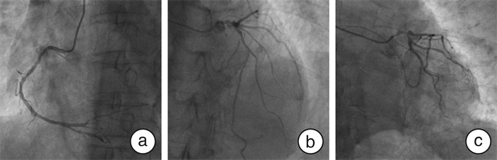
摘要: 经皮冠状动脉介入(percutaneous coronary intervention,PCI)治疗开口病变时常需要支架精确定位。支架突入过多将导致再次介入困难、远期血栓和支架内再狭窄等不良事件发生,但由于开口解剖位置变异、造影剂显影不完全和限制血流等原因,常导致近端支架边缘位置无法清晰识别。本文报道采用血管内超声(intravascular ultrasound,IVUS)实时定位指导支架定位治疗左主干病变2例,发现IVUS较多体位冠状动脉造影更为高效、精准地识别近端支架落脚点,且显著降低造影剂和放射线用量。
-
关键词:
- 经皮冠状动脉介入治疗 /
- 左主干 /
- 血管内超声 /
- 精确定位
Abstract: The percutaneous coronary intervention(PCI) in coronary ostium lesion often requires precise stent implantation. Excessive stent protrusion will lead to adverse events such as difficulty in re-intervention, long-term thrombosis, and in-stent restenosis. However, due to the variation of the anatomical position of the ostium, incomplete image display, limited blood flow, and so on, the operator is sometimes unable to identify the position of the proximal stent edge during angiography. Here, we used the intravascular ultrasound(IVUS) real-time positioning guided stent implantation for two left main lesion patients, and found that the IVUS real-time guidance is more efficient than multi-position coronary angiography, and more accurately identifies the proximal stent edge, and significantly reduce the dose of contrast medium and radiation. -

-
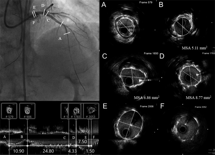
图 5 IVUS指导支架精确定位技术图解
Figure 5. Illustration of the IVUS-guided stent precise implantation technique
图 6 IVUS指导支架精确定位技术体外模式图
Figure 6. The model image of the IVUS-guided stent precise implantation technique
图 11 IVUS指导支架精确定位技术图解
Figure 11. Illustration of the IVUS-guided stent precise implantation technique
-
[1] 陈心怡, 赵国力, 尹德录. 冠状动脉腔内影像学评估斑块性质的研究进展[J]. 临床心血管病杂志, 2023, 39(9): 667-673. https://lcxxg.whuhzzs.com/article/doi/10.13201/j.issn.1001-1439.2023.09.004
[2] Reddy P, Daibes J, Skaf M, et al. The Use of Bumper Wire Technique and Intravascular Ultrasound for Precise Aorto-Ostial Stenting[J]. Front Cardiovasc Med, 2022, 9: 929472. doi: 10.3389/fcvm.2022.929472
[3] 李海蓬, 张健. 血运重建策略对左主干病变所致急性心肌梗死患者预后的影响[J]. 临床心血管病杂志, 2022, 38(4): 303-307. https://lcxxg.whuhzzs.com/article/doi/10.13201/j.issn.1001-1439.2022.04.010
[4] Mavromatis K, Ghazzal Z, Veledar E, et al. Comparison of outcomes of percutaneous coronary intervention of ostial versus nonostial narrowing of the major epicardial coronary arteries[J]. Am J Cardiol, 2004, 94(5): 583-587. doi: 10.1016/j.amjcard.2004.05.020
[5] Freeman M, Clark DJ, Andrianopoulos N, et al. Outcomes after percutaneous coronary intervention of ostial lesions in the era of drug-eluting stents[J]. Catheter Cardiovasc Interv, 2009, 73(6): 763-768. doi: 10.1002/ccd.21941
[6] Kwan TW, James D, Huang Y, et al. Perfection of precise ostial stent placement[J]. J Invasive Cardiol, 2012, 24(7): 354-358.
[7] Dishmon DA, Elhaddi A, Packard K, et al. High incidence of inaccurate stent placement in the treatment of coronary aorto-ostial disease[J]. J Invasive Cardiol, 2011, 23(8): 322-326.
[8] Kern MJ, Ouellette D, Frianeza T. A new technique to anchor stents for exact placement in ostial stenoses: the stent tail wire or Szabo technique[J]. Catheter Cardiovasc Interv, 2006, 68(6): 901-906. doi: 10.1002/ccd.20613
[9] Gutiérrez-Chico JL, Villanueva-Benito I, Villanueva-Montoto L, et al. Szabo technique versus conventional angiographic placement in bifurcations 010-001 of Medina and in aorto-ostial stenting: angiographic and procedural results[J]. EuroIntervention, 2010, 5(7): 801-808. doi: 10.4244/EIJV5I7A134
[10] Vaquerizo B, Serra A, Ormiston J, et al. Bench top evaluation and clinical experience with the Szabo technique: new questions for a complex lesion[J]. Catheter Cardiovasc Interv, 2012, 79(3): 378-389. doi: 10.1002/ccd.23087
[11] Shibata K, Wakabayashi K, Ishinaga T, et al. Feasibility, Safety, and Long-Term Outcomes of Zero-Contrast Percutaneous Coronary Intervention in Patients With Chronic Kidney Disease[J]. Circ J, 2022, 86(5): 787-796. doi: 10.1253/circj.CJ-21-0905
-
 下载:
下载:
图(13)
计量
- 文章访问数: 1214
- PDF下载数: 250
- 施引文献: 0