
A case report of left ventricular lead re-implantation assisted by balloon dilation technique after tricuspid valve replacement
-
摘要: 本病例为既往行三尖瓣金属瓣膜置换术后出现完全性房室传导阻滞经冠状静脉实现DDD起搏模式的患者,4年后出现冠状静脉导线脱位、起搏阈值升高、起搏器电量耗竭。拟行新冠状静脉电极植入过程中发现冠状静脉内存在严重狭窄,通过球囊扩张技术实现了新左心室电极植入。
-
关键词:
- 起搏电极故障 /
- 冠状静脉狭窄 /
- 左心室电极导线再植入 /
- 起搏器更换
Abstract: This case involves a patient who underwent metal tricuspid valve replacement and developed complete atrioventricular block, subsequently managed with DDD pacing mode via the coronary sinus. Four years postoperatively, the patient experienced coronary sinus lead dislodgement, increased pacing threshold, and pacemaker battery depletion. During the attempted implantation of a new coronary sinus electrode, severe stenosis within the coronary sinus was discovered. A new left ventricular electrode was successfully implanted with balloon dilation technique. -

-

图 1 患者首次冠状窦电极植入
Figure 1. Initial implantation of the patient's coronary sinus electrode
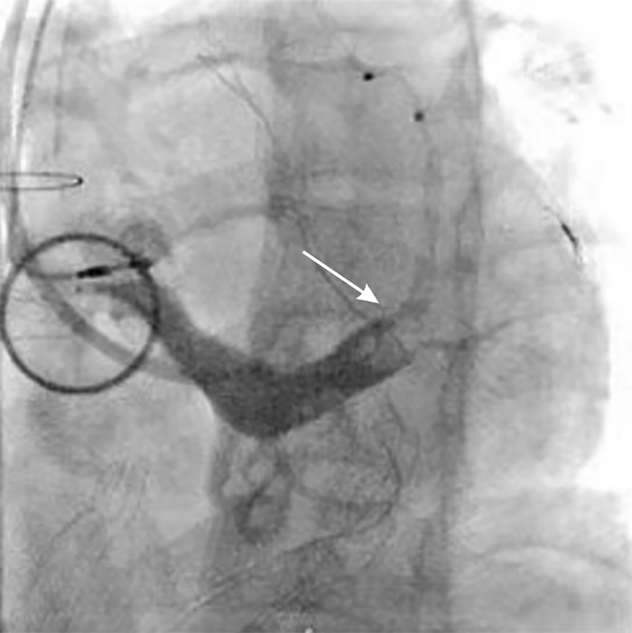
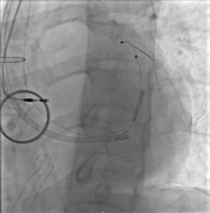
图 6 LAO30°再次行冠状静脉造影可见狭窄部分解除
Figure 6. LAO30° repeat coronary sinus angiography showing resolution of the stenosis
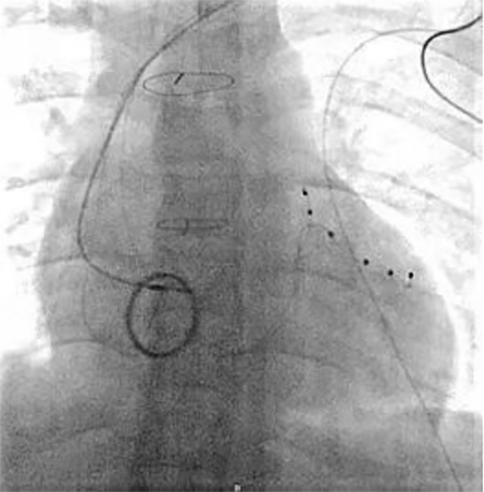
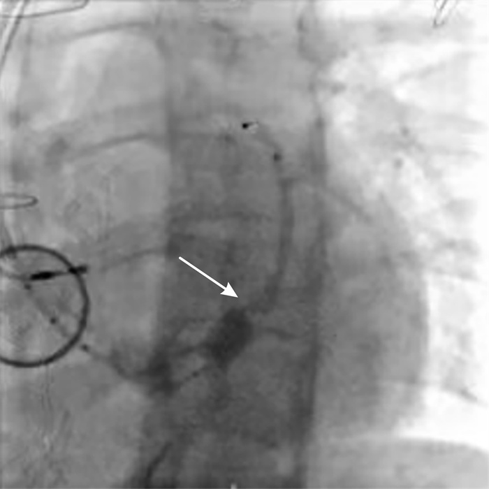
图 7 冠状静脉电极成功置于左室侧静脉(正位)
Figure 7. Coronary vein electrode successfully placed in the left ventricular branch vein(anterior-posterior view)
-
[1] Zheng W, Li D, Li X, et al. Cardiac resynchronization therapy coronary venous left ventricular lead removal and reimplantation: Experience from a single center in China[J]. Exp Ther Med, 2019, 18(3): 2213-2218.
[2] Anderson JH, Mcelhinney DB, Aboulhosn J, et al. Management and Outcomes of Transvenous Pacing Leads in Patients Undergoing Transcatheter Tricuspid Valve Replacement[J]. JACC Cardiovasc Interv, 2020, 13(17): 2012-2020. doi: 10.1016/j.jcin.2020.04.054
[3] Bazire B, Para M, Raffoul R, et al. Prophylactic epicardial pacemaker implantation in tricuspid valve replacement[J]. Eur J Cardiothorac Surg, 2023, 64(6): ezad344. doi: 10.1093/ejcts/ezad344
[4] 乔瀚博, 方潘慧, 陶元, 等. 高龄患者无导线起搏器植入临床疗效观察[J]. 临床心血管病杂志, 2024, 40(7): 570-573. https://lcxxg.whuhzzs.com/article/doi/10.13201/j.issn.1001-1439.2024.07.012
[5] Gao J, Zhang N, Zhang B, et al. A case report of left ventricular lead implantation via total three-dimensional transseptal puncture after tricuspid valve replacement[J]. Front Cardiovasc Med, 2023, 10: 1237967. doi: 10.3389/fcvm.2023.1237967
[6] Glikson M, Nielsen JC, Kronborg MB, et al. 2021 ESC Guidelines on cardiac pacing and cardiac resynchronization therapy[J]. Eur Heart J, 2021, 42(35): 3427-3520. doi: 10.1093/eurheartj/ehab364
[7] Chung MK, Patton KK, Lau CP, et al. 2023 HRS/APHRS/LAHRS guideline on cardiac physiologic pacing for the avoidance and mitigation of heart failure[J]. Heart Rhythm, 2023, 20(9): e17-e91. doi: 10.1016/j.hrthm.2023.03.1538
[8] Lawin D, Stellbrink C. Change in indication for cardiac resynchronization therapy?[J]. Eur J Cardiothorac Surg, 2019, 55(Suppl 1): i11-i16.
[9] Johansen JB, Nielsen JC, Kristensen J, et al. Troubleshooting the difficult left ventricular lead placement in cardiac resynchronization therapy: current status and future perspectives[J]. Expert Rev Med Devices, 2022, 19(4): 341-352. doi: 10.1080/17434440.2022.2075728
[10] Medeiros P, Lousinha A, Oliveira MM. Coronary sinus angioplasty to enable optimal left ventricular lead placement for resynchronization[J]. Heliyon, 2023, 9(6): e16090. doi: 10.1016/j.heliyon.2023.e16090
[11] Gul EE, Abuelatta R, Haseeb S, et al. Venoplasty of a chronic venous occlusion with 'diathermy' for cardiac device lead placement[J]. Indian Pacing Electrophysiol J, 2019, 19(1): 27-29. doi: 10.1016/j.ipej.2018.10.002
[12] Stefanczyk P, Nowosielecka D, Polewczyk A, et al. Safety and Effectiveness of Transvenous Lead Extraction in Patients with Infected Cardiac Resynchronization Therapy Devices; Is It More Risky than Extraction of Other Systems?[J]. Int J Environ Res Public Health, 2022, 19(10): 5803. doi: 10.3390/ijerph19105803
-
 下载:
下载:

图(8)
计量
- 文章访问数: 432
- PDF下载数: 5
- 施引文献: 0