
Clinical application of coronary angioscopy and evaluation of atherosclerotic plaque
-
摘要: 冠状动脉血管镜(coronary angioscopy,CAS)是一种可直接观察心腔内和血管腔表面的独特诊断设备,广泛应用于日本,美国使用甚少,中国少见应用。CAS有助于阐明冠状动脉疾病的病理,可指导血管、先天性心脏病介入治疗,评估介入术后病变恢复情况等。本文回顾近5年来CAS临床应用的相关研究,对其应用范围、评估冠状动脉斑块特征、对比其他腔内影像学的特点以及由CAS确诊的特殊病例进行综述。Abstract: Coronary angioscopy (CAS) is a unique intravascular imaging modality that enables direct visualization of the lumen surfaces of the heart chambers and coronary arteries. While CAS is widely used in Japan, its application remains limited in the United States and is rarely adopted in clinical practice in China. CAS helps to clarify the pathology of coronary artery disease, guides the interventional treatment of vascular and congenital heart disease, and evaluates the recovery after interventional operation. This article reviews clinical research on CAS applications over the past five years, summarizing its scope of use, assessment characteristics for coronary plaque analysis, comparative advantages with other intravascular imaging modalities, and special case diagnoses confirmed through CAS.
-
Key words:
- coronary angioscopy /
- plaque /
- interventional therapy /
- endovascular imaging
-

-
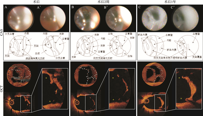
图 1 CAS与OCT评估心肌梗死患者支架植入后状态
Figure 1. Comparison of CAS and OCT for assessing post-stent implantation status in myocardial infarction patients

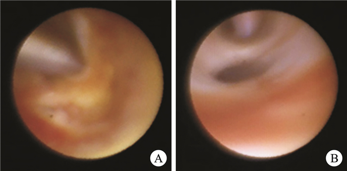
图 2 CAS对比IVUS指导介入治疗
Figure 2. Comparison of CAS and IVUS in guiding interventional therapy
-
[1] Spears JR, Marais HJ, Serur J, et al. In vivo coronary angioscopy[J]. J Am Coll Cardiol, 1983, 1(5): 1311-1314. doi: 10.1016/S0735-1097(83)80145-4
[2] Mitsutake Y, Yano H, Ishihara T, et al. Consensus document on the standard of coronary angioscopy examination and assessment from the Japanese Association of Cardiovascular Intervention and Therapeutics[J]. Cardiovasc Interv Ther, 2022, 37(1): 35-39. doi: 10.1007/s12928-021-00770-x
[3] Nakatani S, Sotomi Y, Otake H, et al. Tissue Characteristics of Stent Coverage Validated by Comparison Between Coronary Angioscopy and Optical Coherence Tomography in Serial Assessment[J]. Circ J, 2023, 87(5): 669. doi: 10.1253/circj.CJ-22-0761
[4] Tsujimura T, Mizote I, Ishihara T, et al. Impact of baseline yellow plaque assessed by coronary angioscopy on vascular response after stent implantation[J]. J Cardiol, 2024, 84(3): 201-207. doi: 10.1016/j.jjcc.2024.04.004
[5] 陈心怡, 赵国力, 尹德录. 冠状动脉腔内影像学评估斑块性质的研究进展[J]. 临床心血管病杂志, 2023, 39(9): 667-673. doi: 10.13201/j.issn.1001-1439.2023.09.004
[6] Itaya N, Tahara N, Bekki M, et al. The role of coronary angioscopy and FDG-PET/CTA in interventional therapy[J]. J Nucl Cardiol, 2023, 30(3): 1269-1271. doi: 10.1007/s12350-022-02915-8
[7] Kim SS, Hijazi ZM, Lang RM, et al. The use of intracardiac echocardiography and other intracardiac imaging tools to guide noncoronary cardiac interventions[J]. J Am Coll Cardiol, 2009, 53(23): 2117-2128. doi: 10.1016/j.jacc.2009.01.071
[8] Miyagawa M, Kojima K, Takahashi K, et al. Association Between Aortic Wall Parameters on Multidetector Computed Tomography and Ruptured Plaques By Nonobstructive General Angioscopy[J]. J Am Heart Assoc, 2024, 13(6): e033233. doi: 10.1161/JAHA.123.033233
[9] Ueda Y, Ohtani T, Shimizu M, et al. Assessment of plaque vulnerability by angioscopic classification of plaque color[J]. Am Heart J, 2004, 148(2): 333-335. doi: 10.1016/j.ahj.2004.03.047
[10] Kimura S, Cho S, Misu Y, et al. Optical coherence tomography and coronary angioscopy assessment of healed coronary plaque components[J]. Int J Cardiovasc Imaging, 2021, 37(10): 2849-2859. doi: 10.1007/s10554-021-02287-z
[11] Omatsu T, Sotomi Y, Kobayashi T, et al. Quantitative Validation of the Coronary Angioscopic Yellow Plaque with Lipid Core Burden Index Assessed by Intracoronary Near-Infrared Spectroscopy[J]. J Atheroscler Thromb, 2022, 29(3): 362-369. doi: 10.5551/jat.60566
[12] Komatsu S, Takahashi S, Takewa M, et al. Demonstrating an Adult Ventricular Septal Defect Using Non-obstructive General Angioscopy[J]. Cureus, 2023, 15(4): e37673.
[13] Yamane H, Ueda Y, Ikeoka K, et al. Case report of a peripheral artery disease patient with its aetiology clarified by retrograde angioscopy[J]. Eur Heart J Case Rep, 2022, 6(10): ytac393. doi: 10.1093/ehjcr/ytac393
[14] Nojima Y, Adachi H, Ihara M, et al. Type Ⅱ Kounis syndrome diagnosed by optical coherence tomography and coronary angioscopy[J]. Cardiol J, 2023, 30(1): 157-158. doi: 10.5603/CJ.2023.0010
[15] Ito S, Tanabe Y, Nawata K, et al. Usefulness of angioscopy for intracardiac tumour biopsy in a patient with malignant lymphoma[J]. Eur Heart J Cardiovasc Imaging, 2023, 24(9): e272. doi: 10.1093/ehjci/jead131
[16] Ueda Y, Matsuo K, Nishimoto Y, et al. In-Stent Yellow Plaque at 1 Year After Implantation Is Associated With Future Event of Very Late Stent Failure: The DESNOTE Study(Detect the Event of Very late Stent Failure From the Drug-Eluting Stent Not Well Covered by Neointima Determined by Angioscopy)[J]. JACC Cardiovasc Interv, 2015, 8(6): 814-821. doi: 10.1016/j.jcin.2014.12.239
[17] Ishihara T, Tsujimura T, Okuno S, et al. Early-and middle-phase arterial repair following bioresorbable-and durable-polymer drug-eluting stent implantation: An angioscopic study[J]. Int J Cardiol, 2019, 285(undefined): 27-31.
[18] Sotomi Y, Suzuki S, Kobayashi T, et al. Impact of the one-year angioscopic findings on long-term clinical events in 504 patients treated with first-generation or second-generation drug-eluting stents: the DESNOTE-X study[J]. Euro Intervention, 2019, 15(7): 631-639.
-
 下载:
下载:

计量
- 文章访问数: 46
- 施引文献: 0