Research on plasma proteome of patients with severe coronary artery calcifications based on mass spectrometry and bioinformatics
-
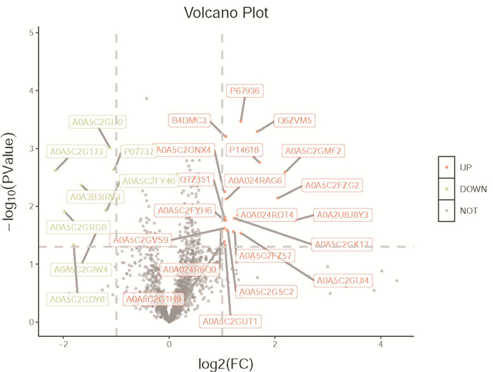
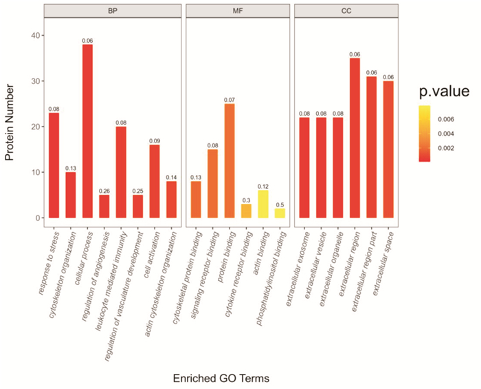
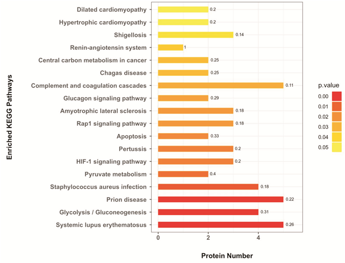
摘要: 目的 基于非标记蛋白组学方法筛选冠状动脉(冠脉)严重钙化病变患者外周血浆中的特异性蛋白质标志物。方法 采用数据非依赖采集质谱技术检测30例冠脉严重钙化病变患者与30例非钙化对照人群血浆,生物信息学软件进一步分析差异表达蛋白质数据。结果 冠脉严重钙化病变患者与非钙化人群血浆相比较,共筛选出表达量差异2倍以上的差异蛋白共28种(其中表达上调的蛋白20种,表达下调的蛋白8种),基因本体论(GO)分析差异表达蛋白主要分布于细胞外区域、细胞外泌体和胞外细胞器部分;生物学过程主要涉及细胞过程、肌动蛋白细胞骨架和应激反应、白细胞介导的免疫和细胞活化;而其分子功能与蛋白质结合、信号受体结合和肌动蛋白结合相关。京都基因与基因组百科全书(KEGG)分析显示差异表达蛋白与补体和凝血级联、糖酵解/糖异生作用、细胞凋亡、HIF-1和Rap1信号通路相关。结论 冠脉严重钙化病变患者和非钙化人群的血浆蛋白质组学存在显著差异,这些差异表达蛋白质有望成为冠脉严重钙化病变鉴别诊断的新型生物标志物。Abstract: Objective Screening of specific plasma protein markers from patients with severe coronary artery calcifications using label free quantitative proteomics.Methods Plasmas were obtained from 30 patients with severe coronary artery calcifications and 30 non-calcification patients and DIA mass spectrometry-based proteomics were used to detect the proteins in plasma. Bioinformatics analysis tools were used to analysis the dysregulated proteomics data.Results A total of 28 plasma proteins were differentially expressed between the coronary calcification patients and controls, with 20 upregulated and 8 downregulated. Gene ontology(GO) analysis indicated that differentially expressed proteins were mainly distributed in extracellular region, extracellular region part, and extracellular organelle. Biological process was mainly involved in cellular process, response to stimulus, actin cytoskeleton organization, leukocyte mediated immunity and cell activation. Molecular function was primarily associated with protein binding, signaling receptor binding, and cytokine receptor binding. Kyoto Encyclopedia of Genes and Genomes(KEGG) analysis revealed that these differentially expressed proteins were mainly involved in complement and coagulation cascade pathway, glycolysis/ gluconeogenesis, Rap1 signaling pathway, HIF-1 signaling pathway, and apoptosis.Conclusion This study demonstrated that significant differences in plasma proteomics were detected between patients with severe coronary artery calcifications and non-calcification controls. These dysregulated proteins would be novel biomarkers for differential diagnosis of severe coronary artery calcification.
-
Key words:
- coronary calcification /
- plasma /
- proteome
-

-
表 1 两组人群基线资料比较
Table 1. General data
X±S 参数 非钙化组(30例) 钙化组(30例) P值 年龄/岁 68.90±1.15 68.37±1.27 0.76 男性/例(%) 25(83.33) 25(83.33) 1.00 BMI/(kg·m-2) 23.88±0.49 23.87±0.67 0.99 高血压病史/例(%) 20(66.67) 21(70.00) 1.00 糖尿病史/例(%) 1(3.33) 3(10.00) 0.61 吸烟史/例(%) 13(43.33) 9(30.00) 0.42 饮酒史/例(%) 11(36.67) 8(26.67) 0.58 收缩压/mmHg 132.60±3.67 137.70±3.68 0.33 舒张压/mmHg 77.60±2.33 79.43±2.06 0.56 空腹血糖/(mmol·L-1) 5.60±0.26 5.51±0.27 0.81 胆固醇/(mmol·L-1) 3.85±0.18 3.92±0.23 0.83 低密度脂蛋白/(mmol·L-1) 2.08±0.15 2.09±0.17 0.96 高密度脂蛋白/(mmol·L-1) 1.08±0.04 1.06±0.05 0.73 甘油三酯/(mmol·L-1) 1.24±0.11 1.35±0.10 0.51 肌酐/(μmol·L-1) 80.29±3.83 74.46±3.87 0.29 表 2 冠脉严重钙化病变患者与非钙化组人群血浆中28种蛋白质的表达差异
Table 2. Differences in the expression of 28 proteins in plasma
编号 蛋白质序列号 蛋白信息描述 基因名称 表达倍数 P值 1 A0A2U8J8Y3 Ig heavy chain variable region IgH 5.201 0.017 2 A0A5C2GMF2 IG c998_light_IGKV4-1_IGKJ2 N/A 4.567 0.003 3 A0A5C2FZG2 IGL c1546_light_IGLV3-21_IGLJ3 N/A 4.147 0.007 4 P14618 Pyruvate kinase PKM PKM 3.273 0.002 5 Q6ZVM5 cDNA FLJ42083 fis,clone TCERX2000613 N/A 3.175 0.001 6 A0A5C2GUI4 IG c1185_light_IGKV1-6_IGKJ1 N/A 2.567 0.029 7 P67936 Tropomyosin alpha-4 chain TPM4 2.560 0.001 8 A0A024RDT4 Lymphocyte cytosolic protein 1(L-plastin),isoform CRA_a LCP1 2.457 0.014 9 A0A5C2GK17 IGH + IGL c357_light_IGKV1D-39_IGKJ1 N/A 2.350 0.016 10 A0A5C2FZ57 IGL c2179_light_IGKV4-1_IGKJ1 N/A 2.321 0.027 11 A0A5C2G5C2 IGH c189_heavy__IGHV3-33_IGHD6-19_IGHJ5 N/A 2.137 0.025 12 A0A024RAG6 Complement C1q subcomponent subunit A ADIC 2.114 0.007 13 B4DMC3 cDNA FLJ60302,highly similar to Neurocan core protein N/A 2.098 0.001 14 A0A5C2FYH6 IGL c2011_light_IGKV1D-33_IGKJ4 N/A 2.081 0.018 15 A0A5C2GUT1 IG c1271_heavy_IGHV3-33_IGHD1-1_IGHJ4 N/A 2.076 0.047 16 Q7Z351 Uncharacterized protein DKFZp686N02209 DKFZp686N02209 2.079 0.016 17 A0A5C2GNX4 IG c870_heavy_IGHV3-66_IGHD3-16_IGHJ1 N/A 2.073 0.005 18 A0A5C2GVS9 IG c657_heavy_IGHV3-7_IGHD5-12_IGHJ4 N/A 2.059 0.024 19 A0A5C2G1H9 IGL c2982_light_IGKV4-1_IGKJ1 N/A 2.047 0.041 20 A0A024R6Q0 HCG2029544,isoform CRA_a hCG_2029544 2.000 0.025 21 P07737 Profilin-1 PFN1 0.483 0.002 22 A0A5C2GIJ0 IG c818_light_IGKV1D-33_IGKJ4 N/A 0.460 0.001 23 A0A5C2FY46 IGL c1564_light_IGKV4-1_IGKJ4 N/A 0.436 0.012 24 A0A5C2GIW4 IGH + IGL c645_heavy_IGHV3-23_IGHD2-21_IGHJ4 N/A 0.384 0.029 25 A0A3B3IRV3 Malignant T-cell-amplified sequence MCTS2P 0.316 0.004 26 A0A5C2GDY8 IGH + IGL c247_heavy_IGHV3-48_IGHD3-9_IGHJ6 N/A 0.287 0.041 27 A0A5C2GRG0 IG c1788_heavy_IGHV1-46_IGHD5-18_IGHJ3 N/A 0.254 0.012 28 A0A5C2G173 IGL c2300_light_IGKV1D-13_IGKJ4 N/A 0.226 0.002 N/A=not avaible(未提供)。 -
[1] Lee SJ, Lee IK, Jeon JH. vascular calcification-new insights into its mechanism[J]. Int J Mol Sci, 2020, 21(8): 2685. doi: 10.3390/ijms21082685
[2] Pino LK, Rose J, O'Broin A, et al. Emerging mass spectrometry-based proteomics methodologies for novel biomedical applications[J]. Biochem Soc Trans, 2020, 48(5): 1953-1966. doi: 10.1042/BST20191091
[3] Xu LL, Young A, Zhou A, et al. Machine learning in mass spectrometric analysis of DIA Data[J]. Proteomics, 2020, 20(21-22): e1900352. doi: 10.1002/pmic.201900352
[4] Scanlon PJ, Faxon DP, Audet AM, et al. ACC/AHA guidelines for coronary angiography[J]. J Am Coll Cardiol, 1999, 33(6): 1756-1824. doi: 10.1016/S0735-1097(99)00126-6
[5] 王莽原, 宋雷, 霍强, 等. 蛋白质组学临床转化研究主要进展及在心脏研究领域应用的挑战和展望[J]. 中国循环杂志, 2021, 36(4): 412-416. https://www.cnki.com.cn/Article/CJFDTOTAL-ZGXH202104018.htm
[6] Duan M, Zhao WL, Zhou L, et al. Omics research in vascular calcification[J]. Clin Chim Acta, 2020, 511: 319-328. doi: 10.1016/j.cca.2020.10.022
[7] Brunner PM, He H, Pavel AB, et al. The blood proteomic signature of early-onset pediatric atopic dermatitis shows systemic inflammation and is distinct from adult long-standing disease[J]. J Am Acad Dermatol, 2019, 81(2): 510-519. doi: 10.1016/j.jaad.2019.04.036
[8] Murota A, Suzuki K, Kassai Y, et al. Serum proteomic analysis identifies interleukin 16 as a biomarker for clinical response during early treatment of rheumatoid arthritis[J]. Cytokine, 2016, 78: 87-93. doi: 10.1016/j.cyto.2015.12.002
[9] Ma WQ, Sun XJ, Zhu Y, et al. PDK4 promotes vascular calcification by interfering with autophagic activity and metabolic reprogramming[J]. Cell Death Dis, 2020, 11(11): 991. doi: 10.1038/s41419-020-03162-w
[10] 刘佳佳, 曹宇, 盛喆. 冠状动脉钙化的病理生理机制及钙化评估进展[J]. 临床心血管病杂志, 2020, 36(8): 768-772. https://www.cnki.com.cn/Article/CJFDTOTAL-LCXB202008019.htm
[11] Martínez-López D, Roldan-Montero R, García-Marqués F, et al. Complement C5 Protein as a Marker of Subclinical Atherosclerosis[J]. J Am Coll Cardiol, 2020, 75(16): 1926-1941. doi: 10.1016/j.jacc.2020.02.058
[12] Haskard DO, Boyle JJ, Mason JC. The role of complement in atherosclerosis[J]. Curr Opin Lipidol, 2008, 19(5): 478-482. doi: 10.1097/MOL.0b013e32830f4a06
[13] Snyder ML, Shields KJ, Korytkowski MT, et al. Complement protein C3 and coronary artery calcium in middle-aged women with polycystic ovary syndrome and controls[J]. Gynecol Endocrinol, 2014, 30(7): 511-515. doi: 10.3109/09513590.2014.895985
[14] Wang Y, Miao Y, Gong K, et al. Plasma complement protein C3a level was associated with abdominal aortic calcification in patients on hemodialysis[J]. J Cardiovasc Transl Res, 2019, 12(5): 496-505. doi: 10.1007/s12265-019-09885-2
[15] Allison SJ. Hypertension: Complement C1 and β-catenin in hypertensive arterial remodelling[J]. Nat Rev Nephrol, 2015, 11(5): 255.
[16] 匡圆圆, 马琦琳, 张赛丹. 血清补体C1q水平与老年患者冠状动脉狭窄程度的相关性研究[J]. 临床心血管病杂志, 2018, 34(11): 1077-1080. https://www.cnki.com.cn/Article/CJFDTOTAL-LCXB201811010.htm
[17] Shi J, Yang Y, Cheng A, et al. Metabolism of vascular smooth muscle cells in vascular diseases[J]. Am J Physiol Heart Circ Physiol, 2020, 319(3): H613-H631. doi: 10.1152/ajpheart.00220.2020
[18] Zhu Y, Ji JJ, Wang XD, et al. Periostin promotes arterial calcification through PPARγ-related glucose metabolism reprogramming[J]. Am J Physiol Heart Circ Physiol, 2021, 320(6): H2222-H2239. doi: 10.1152/ajpheart.01009.2020
[19] Phadwal K, Vrahnas C, Ganley IG, et al. Mitochondrial dysfunction: cause or consequence of vascular calcification?[J]. Front Cell Dev Biol, 2021, 9: 611922. doi: 10.3389/fcell.2021.611922
[20] Mokas S, Larivière R, Lamalice L, et al. Hypoxia-inducible factor-1 plays a role in phosphate-induced vascular smooth muscle cell calcification[J]. Kidney Int, 2016, 90(3): 598-609. doi: 10.1016/j.kint.2016.05.020
[21] Martinez-Val A, Bekker-Jensen DB, Hogrebe A, et al. Data processing and analysis for DIA-based phosphoproteomics using spectronaut[J]. MethodsMol Biol, 2021, 2361: 95-107.
-


 下载:
下载:


