
-
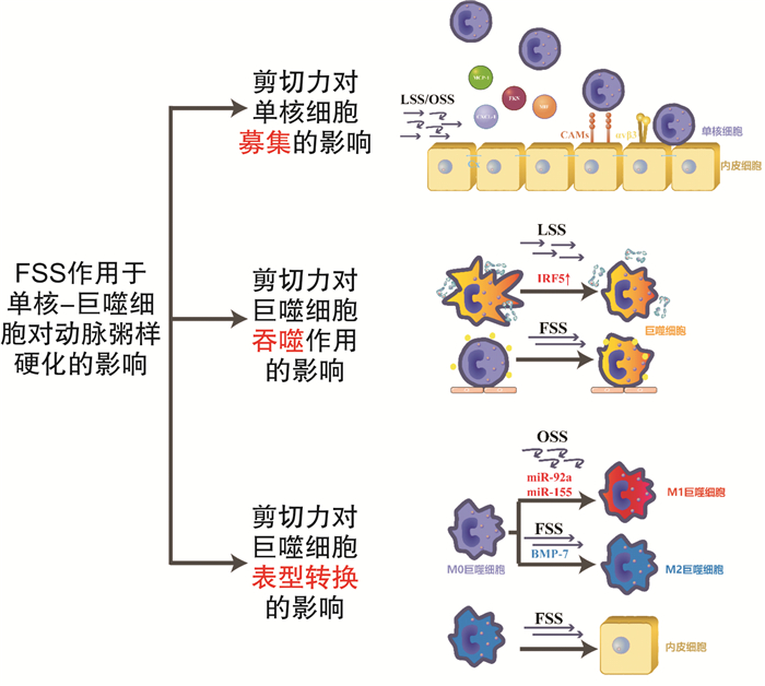
摘要: 流体剪切应力的机械力学刺激在生物体内转化为生物化学信号,该信号可调节单核-巨噬细胞的表型和功能,如招募单核细胞、影响巨噬细胞吞噬功能和细胞极化方向等,并参与了动脉粥样硬化过程。本文对近年研究进展中流体剪切力作用于单核巨噬细胞的影响和机制作一综述。Abstract: The mechanical stimulation of fluid shear stress is transduced into biochemical signals in organisms that regulate the phenotype and functions of monocytes/macrophages. These signals have influences on monocyte recruitment, macrophage phagocytosis and polarization. Then, they participate in process of atherosclerosis. This review covers recent advances in the effects and mechanisms of fluid shear stress on monocytes/macrophages.
-
Key words:
- fluid shear stress /
- monocytes /
- macrophages /
- atherosclerosis
-

-
[1] Gijsen F, Katagiri Y, Barlis P, et al. Expert recommendations on the assessment of wall shear stress in human coronary arteries: existing methodologies, technical considerations, and clinical applications[J]. Eur Heart J, 2019, 40(41): 3421-3433. doi: 10.1093/eurheartj/ehz551
[2] Tabas I, Bornfeldt KE. Macrophage phenotype and function in different stages of atherosclerosis[J]. Circ Res, 2016, 118(4): 653-667. doi: 10.1161/CIRCRESAHA.115.306256
[3] Friedman MH, Deters OJ, Mark FF, et al. Arterial geometry affects hemodynamics. A potential risk factor for athersoclerosis[J]. Atherosclerosis, 1983, 46(2): 225-231. doi: 10.1016/0021-9150(83)90113-2
[4] Tarbell JM. Mass transport in arteries and the localization of atherosclerosis[J]. Annu Rev Biomed Eng, 2003, 5: 79-118. doi: 10.1146/annurev.bioeng.5.040202.121529
[5] Baeyens N, Bandyopadhyay C, Coon BG, et al. Endothelial fluid shear stress sensing in vascular health and disease[J]. J Clin Invest, 2016, 126(3): 821-828. doi: 10.1172/JCI83083
[6] 杨俊杰, 杨晓波, 荆晶, 等. 冠状动脉粥样硬化病变流体力学无创评估的初步研究[J]. 中华心血管病杂志, 2017, 45(8): 716-721. doi: 10.3760/cma.j.issn.0253-3758.2017.08.019
[7] De Wilde D, Trachet B, De Meyer GRY, et al. Shear stress metrics and their relation to atherosclerosis: an in vivo follow-up study in atherosclerotic mice[J]. Ann Biomed Eng, 2016, 44(8): 2327-2338. doi: 10.1007/s10439-015-1540-z
[8] Cheng C, Tempel D, Van Haperen R, et al. Atherosclerotic lesion size and vulnerability are determined by patterns of fluid shear stress[J]. Circulation, 2006, 113(23): 2744-2753. doi: 10.1161/CIRCULATIONAHA.105.590018
[9] Xing R, Moerman AM, Ridwan Y, et al. Temporal and spatial changes in wall shear stress during atherosclerotic plaque progression in mice[J]. R Soc Open Sci, 2018, 5(3): 171447. doi: 10.1098/rsos.171447
[10] Varela A, Piperi C, Sigala F, et al. Elevated expression of mechanosensory polycystins in human carotid atherosclerotic plaques: association with p53 activation and disease severity[J]. Sci Rep, 2015, 5: 13461. doi: 10.1038/srep13461
[11] Qiao R, Qiao H, Zhang Y, et al. Molecular imaging of vulnerable atherosclerotic plaques in vivo with osteopontin-specific upconversion nanoprobes[J]. ACS Nano, 2017, 11(2): 1816-1825. doi: 10.1021/acsnano.6b07842
[12] Baeriswyl DC, Prionisti I, Peach T, et al. Disturbed flow induces a sustained, stochastic NF-κB activation which may support intracranial aneurysm growth in vivo[J]. Sci Rep, 2019, 9(1): 4738. doi: 10.1038/s41598-019-40959-y
[13] Aoki T, Yamamoto K, Fukuda M, et al. Sustained expression of MCP-1 by low wall shear stress loading concomitant with turbulent flow on endothelial cells of intracranial aneurysm[J]. Acta Neuropathol Commun, 2016, 4(1): 48. doi: 10.1186/s40478-016-0318-3
[14] Aoki T, Frösen J, Fukuda M, et al. Prostaglandin E2-EP2-NF-κB signaling in macrophages as a potential therapeutic target for intracranial aneurysms[J]. Sci Signal, 2017, 10(465): 100.
[15] Weinbaum S, Tarbell JM, Damiano ER. The structure and function of the endothelial glycocalyx layer[J]. Annu Rev Biomed Eng, 2007, 9: 121-167. doi: 10.1146/annurev.bioeng.9.060906.151959
[16] Cancel LM, Ebong EE, Mensah S, et al. Endothelial glycocalyx, apoptosis and inflammation in an atherosclerotic mouse model[J]. Atherosclerosis, 2016, 252: 136-146. doi: 10.1016/j.atherosclerosis.2016.07.930
[17] Zhang J, Kong X, Wang Z, et al. AMP-activated protein kinase regulates glycocalyx impairment and macrophage recruitment in response to low shear stress[J]. FASEB J, 2019, 33(6): 7202-7212. doi: 10.1096/fj.201801869RRR
[18] Cooper S, Teoh H, Campeau MA, et al. Empagliflozin restores the integrity of the endothelial glycocalyx in vitro[J]. Mol Cell Biochem, 2019, 459(1-2): 121-130. doi: 10.1007/s11010-019-03555-2
[19] Chen J, Green J, Yurdagul A, et al. αvβ3 Integrins mediate flow-induced NF-κB activation, proinflammatory gene expression, and early atherogenic inflammation[J]. Am J Pathol, 2015, 185(9): 2575-2589. doi: 10.1016/j.ajpath.2015.05.013
[20] Wong CW, Christen T, Roth I, et al. Connexin 37 protects against atherosclerosis by regulating monocyte adhesion[J]. Nat Med, 2006, 12(8): 950-954. doi: 10.1038/nm1441
[21] Pfenniger A, Meens MJ, Pedrigi RM, et al. Shear stress-induced atherosclerotic plaque composition in ApoE(-/-)mice is modulated by connexin37[J]. Atherosclerosis, 2015, 243(1): 1-10. doi: 10.1016/j.atherosclerosis.2015.08.029
[22] Cheng C, Tempel D, Van Haperen R, et al. Shear stress-induced changes in atherosclerotic plaque composition are modulated by chemokines[J]. J Clin Invest, 2007, 117(3): 616-626. doi: 10.1172/JCI28180
[23] Dekker RJ, Van Thienen JV, Rohlena J, et al. Endothelial KLF2 links local arterial shear stress levels to the expression of vascular tone-regulating genes[J]. Am J Pathol, 2005, 167(2): 609-618. doi: 10.1016/S0002-9440(10)63002-7
[24] Qiao C, Li S, Lu H, et al. Laminar flow attenuates macrophage migration inhibitory factor expression in endothelial cells[J]. Sci Rep, 2018, 8(1): 2360. doi: 10.1038/s41598-018-20885-1
[25] Sunderkötter C, Goebeler M, Schulze-Osthoff K, et al. Macrophage-derived angiogenesis factors[J]. Pharmacol Ther, 1991, 51(2): 195-216. doi: 10.1016/0163-7258(91)90077-Y
[26] 吴金锟, 马树沛, 孙玲玲, 等. 弥漫大B细胞淋巴瘤微环境中CD68表达与微血管密度的关系及意义[J]. 临床血液学杂志, 2019, 32(3): 193-196.
[27] Fujita M, Sasayama S. Coronary collateral growth and its therapeutic application to coronary artery disease[J]. Circ J, 2010, 74(7): 1283-1289. doi: 10.1253/circj.CJ-10-0376
[28] Vries MH, Wagenaar A, Verbruggen SE, et al. CXCL1 promotes arteriogenesis through enhanced monocyte recruitment into the peri-collateral space[J]. Angiogenesis, 2015, 18(2): 163-171. doi: 10.1007/s10456-014-9454-1
[29] Zhu H, Zhang M, Liu Z, et al. AMP-activated protein Kinase α1 in macrophages promotes collateral remodeling and arteriogenesis in mice in vivo[J]. Arterioscler Thromb Vasc Biol, 2016, 36(9): 1868-1878. doi: 10.1161/ATVBAHA.116.307743
[30] Heuslein JL, Meisner JK, Li X, et al. Mechanisms of amplified arteriogenesis in collateral artery segments exposed to reversed flow direction[J]. Arterioscler Thromb Vasc Biol, 2015, 35(11): 2354-2365. doi: 10.1161/ATVBAHA.115.305775
[31] Seneviratne AN, Edsfeldt A, Cole JE, et al. Interferon regulatory factor 5 controls necrotic core formation in atherosclerotic lesions by impairing efferocytosis[J]. Circulation, 2017, 136(12): 1140-1154. doi: 10.1161/CIRCULATIONAHA.117.027844
[32] Moore TL, Hauser D, Gruber T, et al. Cellular shuttles: monocytes/macrophages exhibit transendothelial transport of nanoparticles under physiological flow[J]. ACS Appl Mater Interfaces, 2017, 9(22): 18501-18511. doi: 10.1021/acsami.7b03479
[33] Seneviratne AN, Cole JE, Goddard ME, et al. Low shear stress induces M1 macrophage polarization in murine thin-cap atherosclerotic plaques[J]. J Mol Cell Cardiol, 2015, 89(Pt B): 168-172.
[34] Lu Q, Meng Q, Qi M, et al. Shear-sensitive lncRNA AF131217.1 inhibits inflammation in HUVECs via regulation of KLF4[J]. Hypertension, 2019, 73(5): e25-e34.
[35] Chang YJ, Li YS, Wu CC, et al. Extracellular microRNA-92a mediates endothelial cell-macrophage communication[J]. Arterioscler Thromb Vasc Biol, 2019, 39(12): 2492-2504. doi: 10.1161/ATVBAHA.119.312707
[36] Li Z, Martin M, Zhang J, et al. Krüppel-like factor 4 regulation of cholesterol-25-hydroxylase and liver X receptor mitigates atherosclerosis susceptibility[J]. Circulation, 2017, 136(14): 1315-1330. doi: 10.1161/CIRCULATIONAHA.117.027462
[37] 杨朋康, 董昕. 血浆微小核糖核酸-155表达与冠状动脉慢血流的相关性研究[J]. 临床血液学杂志, 2020, 33(6): 394-398.
[38] Nazari-Jahantigh M, Wei Y, Noels H, et al. MicroRNA-155 promotes atherosclerosis by repressing Bcl6 in macrophages[J]. J Clin Invest, 2012, 122(11): 4190-4202. doi: 10.1172/JCI61716
[39] Teng C, Lin C, Huang F, et al. Intracellular codelivery of anti-inflammatory drug and anti-miR 155 to treat inflammatory disease[J]. Acta Pharm Sin B, 2020, 10(8): 1521-1533. doi: 10.1016/j.apsb.2020.06.005
[40] Cheng HS, Besla R, Li A, et al. Paradoxical suppression of atherosclerosis in the absence of microRNA-146a[J]. Circ Res, 2017, 121(4): 354-367. doi: 10.1161/CIRCRESAHA.116.310529
[41] Li K, Ching D, Luk FS, et al. Apolipoprotein E enhances microRNA-146a in monocytes and macrophages to suppress nuclear factor-κB-driven inflammation and atherosclerosis[J]. Circ Res, 2015, 117(1): e1-e11. doi: 10.1161/RES.0000000000000060
[42] Sharp LA, Lee YW, Goldstein AS. Effect of low-frequency pulsatile flow on expression of osteoblastic genes by bone marrow stromal cells[J]. Ann Biomed Eng, 2009, 37(3): 445-453. doi: 10.1007/s10439-008-9632-7
[43] Shoulders H, Garner KH, Singla DK. Macrophage depletion by clodronate attenuates bone morphogenetic protein-7 induced M2 macrophage differentiation and improved systolic blood velocity in atherosclerosis[J]. Transl Res, 2019, 203: 1-14. doi: 10.1016/j.trsl.2018.07.006
[44] Wissing TB, Van Haaften EE, Koch SE, et al. Hemodynamic loads distinctively impact the secretory profile of biomaterial-activated macrophages-implications for in situ vascular tissue engineering[J]. Biomater Sci, 2019, 8(1): 132-147.
[45] Smith RJ, Nasiri B, Kann J, et al. Endothelialization of arterial vascular grafts by circulating monocytes[J]. Nat Commun, 2020, 11(1): 1622. doi: 10.1038/s41467-020-15361-2
[46] Wang L, Luo JY, Li B, et al. Integrin-YAP/TAZ-JNK cascade mediates atheroprotective effect of unidirectional shear flow[J]. Nature, 2016, 540(7634): 579-582. doi: 10.1038/nature20602
[47] Seo Y, Park J, Choi W, et al. Antiatherogenic effect of resveratrol attributed to decreased expression of ICAM-1(Intercellular Adhesion Molecule-1)[J]. Arterioscler Thromb Vasc Biol, 2019, 39(4): 675-684. doi: 10.1161/ATVBAHA.118.312201
[48] Nallasamy P, Si H, Babu PV, et al. Sulforaphane reduces vascular inflammation in mice and prevents TNF-α-induced monocyte adhesion to primary endothelial cells through interfering with the NF-κB pathway[J]. J Nutr Biochem, 2014, 25(8): 824-833. doi: 10.1016/j.jnutbio.2014.03.011
[49] Nicol ED, Norgaard BL, Blanke P, et al. The future of cardiovascular computed tomography: advanced analytics and clinical insights[J]. JACC Cardiovasc Imaging, 2019, 12(6): 1058-1072. doi: 10.1016/j.jcmg.2018.11.037
[50] Tong W, Hui H, Shang W, et al. Highly sensitive magnetic particle imaging of vulnerable atherosclerotic plaque with active myeloperoxidase-targeted nanoparticles[J]. Theranostics, 2021, 11(2): 506-521. doi: 10.7150/thno.49812
[51] Becher T, Riascos-Bernal DF, Kramer DJ, et al. Three-dimensional imaging provides detailed atherosclerotic plaque morphology and reveals angiogenesis after carotid artery ligation[J]. Circ Res, 2020, 126(5): 619-632. doi: 10.1161/CIRCRESAHA.119.315804
[52] Dazzi M, Rowland EM, Mohri Z, et al. 3D confocal microscope imaging of macromolecule uptake in the intact brachiocephalic artery[J]. Atherosclerosis, 2020, 310: 93-101. doi: 10.1016/j.atherosclerosis.2020.07.002
[53] 《中国冠状动脉血流储备分数测定技术临床路径专家共识》专家组. 中国冠状动脉血流储备分数测定技术临床路径专家共识[J]. 中国介入心脏病学杂志, 2019, 27(3): 121-133. doi: 10.3969/j.issn.1004-8812.2019.03.001
[54] Tian F, Yu W, Huang J, et al. First presentation of integration of intravascular optical coherence tomography and computational fractional flow reserve[J]. Int J Cardiovasc Imaging, 2019, 35(4): 601-602. doi: 10.1007/s10554-018-1491-1
-
 下载:
下载:
图(1)
计量
- 文章访问数: 3093
- PDF下载数: 2585
- 施引文献: 0