
-
摘要: 几十年来,尽管心力衰竭(心衰)的治疗取得了相当大的进展,但心衰依然是全世界最主要的死亡原因之一。绝大多数能够诱发心衰的心血管疾病受到遗传和环境因素影响,基础及转化研究领域的最新研究进展,如遗传分析和单细胞分析,有助于揭示心衰的发病机制或病理生理机制,有望在心衰的诊断和预后分层方面发挥作用,为开发新的治疗手段提供新的思路和方法。本文主要阐述心衰转化研究方面的最新进展。Abstract: Despite significant progress in the treatment of heart failure over the past few decades, heart failure remains one of the leading causes of death worldwide. Most cardiovascular diseases that predispose individuals to heart failure are caused by genetic and environmental factors. Recent advances in basic and translational research fields, such as genomic analysis and single-cell analysis, has shown great potential for unveiling the pathogenesis and/or pathophysiology, and can contribute to the diagnosis and prognostic stratification of heart failure, which provide new ideas and methods for the development of novel treatment methods. Here, we summarize the recent advances in translational research on heart failure.
-
Key words:
- heart failure /
- translational research /
- genomic analysis /
- single-cell analysis
-

-
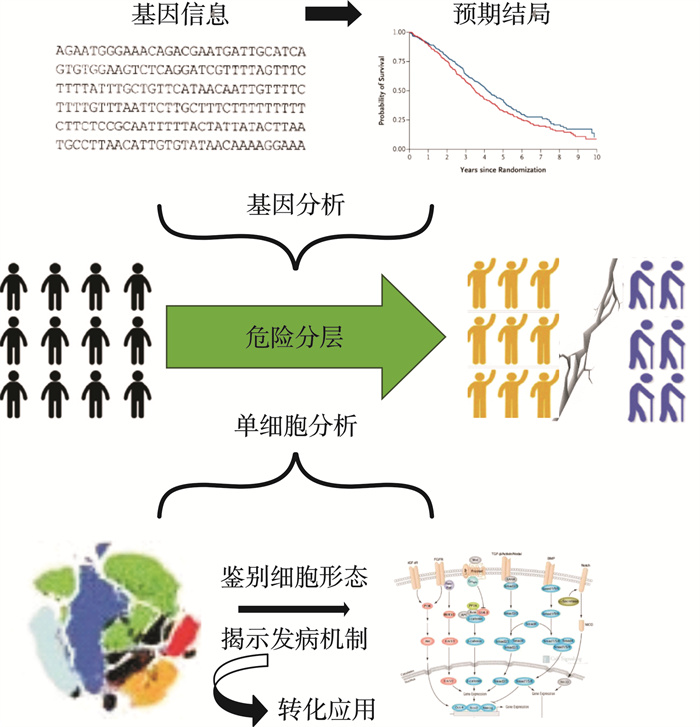
图 1 心衰诊断及预后分层的转化研究
Figure 1. Translational research on diagnosis and prognostic stratification of heart failure
表 1 近期关于心衰scRNA-seq或snRNA-seq分析的研究
Table 1. Recent studies on scRNA-seq and snRNA-seq analysis in heart failure
小鼠 年份 方法 样本 手术 2017[33] snRNA-seq 左心室CMs 对照组、TAC 2018[15] scRNA-seq 左心室CMs 对照组、TAC 2019[34] scRNA-seq 左心室CMs 对照组、TAC 2019[35] scRNA-seq 整个心脏的CD45+细胞 对照组、TAC 2020[16] scRNA-seq 左心室CMs 对照组、TAC 2020[36] scRNA-seq 左心室CMs 对照组、TAC 2022[17] scRNA-seq 左心室CMs 对照组、TAC、心肌梗死组 人类 年份 方法 样本 患者 2017[33] snRNA-seq 左心室CMs 健康组、扩张型心肌病组 2018[15] scRNA-seq 左心室CMs 健康组、扩张型心肌病组 2020[16] scRNA-seq 左心室CMs 对照组、TAC 2020[37] scRNA-seq 左心室或心房的细胞 健康组、心衰组、部分因植入左心室辅助装置而恢复 2021[38] scRNA-seq、T细胞受体测序 左心室或右心室的细胞 健康组、扩张型心肌病组、缺血性心肌病组 2022[39] scRNA-seq 左心室细胞核 健康组、扩张性型心肌病组、肥厚性心肌病组 2022[17] scRNA-seq 左心室CMs 健康组、扩张型心肌病组 2022[40] snRNA-seq 左心室细胞核 健康组、扩张型心肌病组、致心律失常性心肌病组 2022[41] scRNA-seq、snRNA-seq 左心室CMs 健康组、扩张型心肌病组 TAC:主动脉弓缩窄模型;CMs:心肌细胞。  下载: 导出CSV
-
[1] Mosterd A, Hoes AW. Clinical epidemiology of heart failure[J]. Heart, 2007, 93(9): 1137-1146. doi: 10.1136/hrt.2003.025270
[2] 国家心血管病医疗质量控制中心专家委员会心力衰竭专家工作组. 2020中国心力衰竭医疗质量控制报告[J]. 中国循环杂志, 2021, 36(3): 221-238.
[3] Virani SS, Alonso A, Aparicio HJ, et al. Heart disease and stroke statistics—2021 update: a report from the american heart association[J]. Circulation, 2021, 143(8): 254-743.
[4] Tomomi I, Kaku H, Shouji M, et al. Clinical characteristics and outcomes of hospitalized patients with heart failure from the large-scale japanese registry of acute decompensated heart failure(JROADHF)[J]. Circ J, 2021, 85(9): 1438-1450. doi: 10.1253/circj.CJ-20-0947
[5] 中华医学会, 中华医学会杂志社, 中华医学会全科医学分会, 等. 中国心力衰竭基层诊疗与管理指南(2024年)[J]. 中华全科医师杂志, 2024, 23(6): 549-577.
[6] Yaku H, Ozasa N, Morimoto T, et al. Demographics, management, and in-hospital outcome of hospitalized acute heart failure syndrome patients in contemporary real clinical practice in japan ― observations from the prospective, multicenter kyoto congestive heart failure(KCHF)registry[J]. Circ J, 2018, 82(11): 2811-2819. doi: 10.1253/circj.CJ-17-1386
[7] Crespo-Leiro MG, Anker SD, Maggioni AP, et al. European society of cardiology heart failure long-term registry(ESC-HF-LT): 1-year follow-up outcomes and differences across regions[J]. Eur J Heart Fail, 2016, 18(6): 613-625. doi: 10.1002/ejhf.566
[8] 王建昌, 刘平, 陈力达, 等. 老年人和中青年人首发急性左心衰竭诱因和病因的对比研究[J]. 中国老年学杂志, 2006, 26(12): 1607-1608.
[9] Okura Y, Ramadan MM, Ohno Y, et al. Impending epidemic: future projection of heart failure in japan to the year 2055[J]. Circ J, 2008, 72(3): 489-491. doi: 10.1253/circj.72.489
[10] Yasuda S, Miyamoto Y, Ogawa H. Current status of cardiovascular medicine in the aging society of Japan[J]. Circulation, 2018, 138(10): 965-967. doi: 10.1161/CIRCULATIONAHA.118.035858
[11] Hamaguchi S, Kinugawa S, Sobirin M, et al. Mode of death in patients with heart failure and reduced vs. preserved ejection fraction: report from the registry of hospitalized heart failure patients[J]. Circ J, 2012, 76(7): 1662-1629.
[12] Kaichi R, Marume K, Nakai M, et al. Relationship between heart failure hospitalization costs and left ventricular ejection fraction in an advanced aging society[J]. Circ Rep, 2022, 4(1): 48-58. doi: 10.1253/circrep.CR-21-0134
[13] Olchanski N, Vest AR, Cohen JT, et al. Cost comparison across heart failure patients with reduced and preserved ejection fractions: analyses of inpatient decompensated heart failure admissions[J]. Int J Cardiol, 2018, 261: 103-108. doi: 10.1016/j.ijcard.2018.03.024
[14] Suzuki A, Shiga T, Kawashiro N, et al. Changes in characteristics and outcomes in Japanese patients with heart failure from the 2000 s to the 2010 s: the HIJ-HF cohorts[J]. J Cardiol, 2020, 76(2): 132-138. doi: 10.1016/j.jjcc.2020.02.008
[15] Nomura S, Satoh M, Fujita T, et al. Cardiomyocyte gene programs encoding morphological and functional signatures in cardiac hypertrophy and failure[J]. Nat Commun, 2018, 9(1): 4435. doi: 10.1038/s41467-018-06639-7
[16] Yamaguchi T, Sumida TS, Nomura S, et al. Cardiac dopamine d1 receptor triggers ventricular arrhythmia in chronic heart failure[J]. Nat Commun, 2020, 11(1): 4364-4364. doi: 10.1038/s41467-020-18128-x
[17] Ko T, Nomura S, Yamada S, et al. Cardiac fibroblasts regulate the development of heart failure via Htra3-TGF-β-IGFBP7 axis[J]. Nat Commun, 2022, 13(1): 3275-3275. doi: 10.1038/s41467-022-30630-y
[18] Ko T, Ffujita K, Nnomura S, et al. Quantification of dna damage in heart tissue as a novel prediction tool for therapeutic prognosis of patients with dilated cardiomyopathy[J]. JACC Basic Transl Sci, 2019, 4(6): 670-680. doi: 10.1016/j.jacbts.2019.05.010
[19] Yamada S, Ko T, Hatsuse S, et al. Spatiotemporal transcriptome analysis reveals critical roles for mechano-sensing genes at the border zone in remodeling after myocardial infarction[J]. Nat Cardiovasc Res, 2022, 1(11): 1072-1083. doi: 10.1038/s44161-022-00140-7
[20] Zhu H, Galdos FX, Lee D, et al. Identification of pathogenic immune cell subsets associated with checkpoint inhibitor-induced myocarditis[J]. Circulation, 2022: 146(4): 316-335. doi: 10.1161/CIRCULATIONAHA.121.056730
[21] Ambale-Venkatesh B, Yang X, Wu CO, et al. Cardiovascular event prediction by machine learning: the multi-ethnic study of atherosclerosis[J]. Circ Res, 2017, 121(9): 1092-1101. doi: 10.1161/CIRCRESAHA.117.311312
[22] Kagiyama N, Piccirilli M, Yanamala N, et al. Machine learning assessment of left ventricular diastolic function based on electrocardiographic features[J]. J Am Coll Cardiol, 2020, 76(8): 930-941. doi: 10.1016/j.jacc.2020.06.061
[23] Lei Y, Tang R, Xu J, et al. Applications of single-cell sequencing in cancer research: progress and perspectives[J]. J Hematol Oncol, 2021, 14(1): 91. doi: 10.1186/s13045-021-01105-2
[24] Yekula A, Tracz J, Rincon-Torroella J, et al. Single-cell rna sequencing of cerebrospinal fluid as an advanced form of liquid biopsy for neurological disorders[J]. Brain Sci, 2022, 12(7): 812-812. doi: 10.3390/brainsci12070812
[25] Green ME, Hiroko W, Anderson LR, et al. A small-molecule inhibitor of sarcomere contractility suppresses hypertrophic cardiomyopathy in mice[J]. Science, 2016, 351(6273): 617-621. doi: 10.1126/science.aad3456
[26] Malik FI, Hartman JJ, Elias KA, et al. Cardiac myosin activation: a potential therapeutic approach for systolic heart failure[J]. Science, 2011, 331(6023): 1439-1443. doi: 10.1126/science.1200113
[27] Najjar SS. Heart failure with preserved ejection fraction[J]. J Am Coll Cardiol, 2009, 54(5): 419-421. doi: 10.1016/j.jacc.2009.05.011
[28] Schiattarella GG, Altamirano F, Tong D, et al. Nitrosative stress drives heart failure with preserved ejection fraction[J]. Nature, 2019, 568(7752): 351-356. doi: 10.1038/s41586-019-1100-z
[29] Tobita T, Nomura S, Fujita T, et al. Genetic basis of cardiomyopathy and the genotypes involved in prognosis and left ventricular reverse remodeling[J]. Sci Rep, 2018, 8(1): 1998. doi: 10.1038/s41598-018-20114-9
[30] Koyama S, Ito K, Terao C, et al. Population-specific and trans-ancestry genome-wide analyses identify distinct and shared genetic risk loci for coronary artery disease[J]. Nat Genet, 2020, 52(11): 1169-1177. doi: 10.1038/s41588-020-0705-3
[31] Yurista SR, Chong CR, Badimon JJ, et al. Therapeutic potential of ketone bodies for patients with cardiovascular disease: JACC State-of-the-Art Review[J]. J Am Coll Cardiol, 2021, 77(13): 1660-1669. doi: 10.1016/j.jacc.2020.12.065
[32] Packer M. Critical reanalysis of the mechanisms underlying the cardiorenal benefits of SGLT2 inhibitors and reaffirmation of the nutrient deprivation signaling/autophagy hypothesis[J]. Circulation, 2022, 146(18): 1383-1405. doi: 10.1161/CIRCULATIONAHA.122.061732
[33] See K, Tan WLW, Lim EH, et al. Single cardiomyocyte nuclear transcriptomes reveal a lincrna-regulated de-differentiation and cell cycle stress-response in vivo[J]. Nat Commun, 2017, 8(1): 225. doi: 10.1038/s41467-017-00319-8
[34] Yekelchyk M, Guenther S, Preussner J, et al. Mono-and multi-nucleated ventricular cardiomyocytes constitute a transcriptionally homogenous cell population[J]. Basic Res Cardiol, 2019, 114(5): 36. doi: 10.1007/s00395-019-0744-z
[35] Martini E, Kunderfranco P, Peano C, et al. Single-cell sequencing of mouse heart immune infiltrate in pressure overload-driven heart failure reveals extent of immune activation[J]. Circulation, 2019, 140(25): 2089-2107. doi: 10.1161/CIRCULATIONAHA.119.041694
[36] Ren Z, Yu P, Li D, et al. Single-cell reconstruction of progression trajectory reveals intervention principles in pathological cardiac hypertrophy[J]. Circulation, 2020, 141(21): 1704-1719. doi: 10.1161/CIRCULATIONAHA.119.043053
[37] Wang L, Yu P, Zhou B, et al. Single-cell reconstruction of the adult human heart during heart failure and recovery reveals the cellular landscape underlying cardiac function[J]. Nat Cell Biol, 2020, 22(1): 108-119. doi: 10.1038/s41556-019-0446-7
[38] Rao M, Wang X, Guo G, et al. Resolving the intertwining of inflammation and fibrosis in human heart failure at single-cell level[J]. Basic Res Cardiol, 2021, 116(1): 55. doi: 10.1007/s00395-021-00897-1
[39] Chaffin M, Papangeli I, Simonson B, et al. Single-nucleus profiling of human dilated and hypertrophic cardiomyopathy[J]. Nature, 2022, 608(7921): 174-180. doi: 10.1038/s41586-022-04817-8
[40] Reichart D, Lindberg EL, Maatz H, et al. Pathogenic variants damage cell composition and single cell transcription in cardiomyopathies[J]. Science, 2022, 377(6606): 1984-1984. doi: 10.1126/science.abo1984
[41] Koenig AL, Shchukina I, Amrute J, et al. Single-cell transcriptomics reveals cell-type-specific diversification in human heart failure[J]. Nat Cardiovasc Res, 2022, 1(3): 263-280. doi: 10.1038/s44161-022-00028-6
-
图(1)
表(1)
计量
- 文章访问数: 936
- PDF下载数: 62
- 施引文献: 0