
Predictive value of coronary CTA in patients with coronary heart disease combined with atrial fibrillation
-
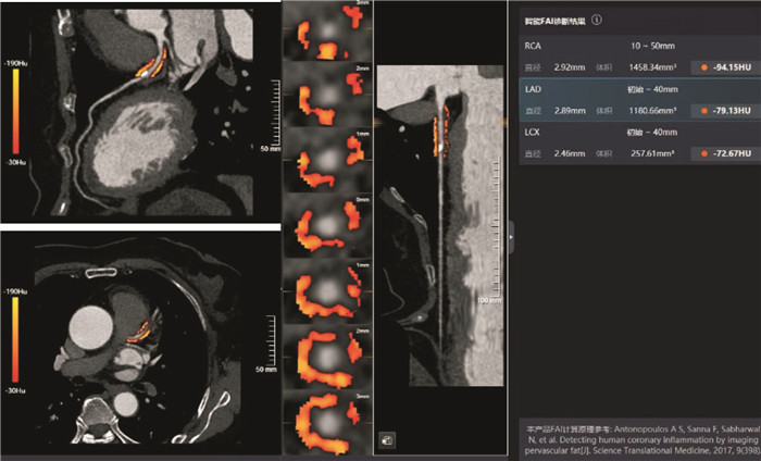
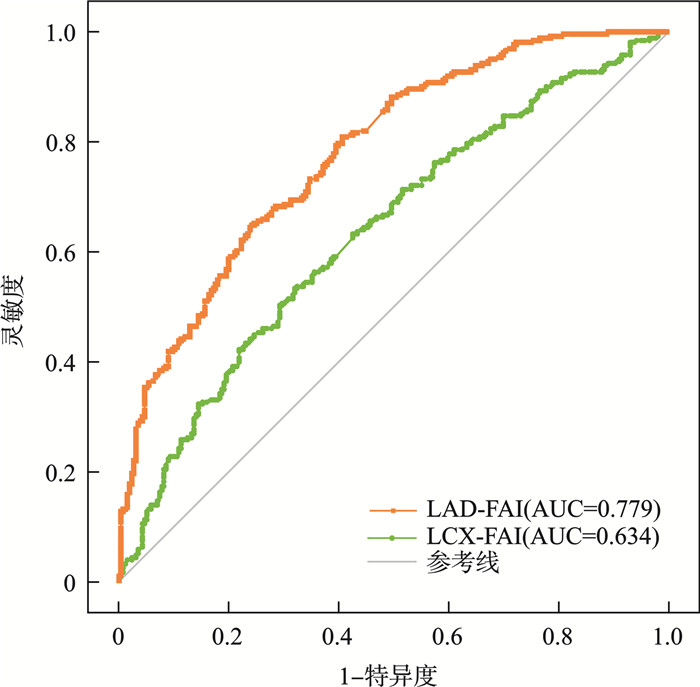
摘要: 目的 探讨冠状动脉(冠脉)周围脂肪衰减指数(FAI)对冠心病(CHD)患者心房颤动(AF)发生的预测价值。 方法 分析2022—2024年于我院入院后确诊CHD的516例患者临床、实验室、心脏超声及冠脉CT造影(CCTA)资料,并测量冠脉周围脂肪组织(PCAT)体积及FAI,随访1年。将所有CHD患者分为AF组255例及对照组261例,对比组间CCTA相关指标。采用单因素和多因素logistic回归分析PCAT参数与CHD患者发生AF的关系,ROC曲线分析其诊断价值。然后分析不同类型AF的PCAT参数的差异。 结果 与对照组比较,AF组入院时LAD-FAI及LCX-FAI较高(P < 0.05),PCAT-V及RCA-FAI两组间无统计学差异。单因素和多因素logistic回归分析后,LAD-FAI及LCX-FAI是AF发生的影响因素(P < 0.05)。LCX-FAI(AUC=0.779)诊断价值高于LAD-FAI(AUC=0.634,P=0.001)。将AF分为持续性房颤(PersAF)与阵发性房颤(PAF),PAF的LCX-FAI低于PersAF(P < 0.05)。 结论 LAD-FAI及LCX-FAI是CHD患者AF事件的独立预测因子,LCX-FAI与AF的类型相关。Abstract: Objective To explore the predictive value of coronary artery fat attenuation index(FAI) for atrial fibrillation(AF) in patients with coronary heart disease(CHD). Methods Clinical, laboratory, cardiac ultrasound, and coronary CT angiography(CCTA) data of 516 patients diagnosed with CHD after admission in our hospital from 2022 to 2024 were analyzed. Pericoronary adipose tissue(PCAT) volume and FAI were measured, and we followed up for 1 year. All CHD patients were divided into an AF group with 255 cases and a control group with 261 cases, and CCTA related indicators were compared between the control groups. Univariate and multivariate logistic regression were used to analyze the relationship between PCAT parameters and the occurrence of AF in CHD patients, and ROC curve was used to analyze its diagnostic value. Then analyze the differences in PCAT parameters for different types of AF. Results Compared with the control group, the AF group had higher levels of LAD-FAI and LCX-FAI upon admission(P < 0.05), while there was no statistically significant difference between the PCAT-V and RCA-FAI groups. In univariate and multivariate logistic regression analysis, LAD-FAI and LCX-FAI were identified as influencing factors for the occurrence of atrial fibrillation(P < 0.05). The diagnostic value of LCX-FAI(AUC=0.779) is higher than that of LAD-FAI(AUC=0.634, P=0.001). AF is divided into persistent atrial fibrillation(PersAF) and paroxysmal atrial fibrillation(PAF), and PAF having a lower LCX-FAI than PersAF(P < 0.05). Conclusion LAD-FAI and LCX-FAI are independent predictors of AF in CHD patients, and LCX-FAI is associated with the type of AF.
-
Key words:
- pericoronary adipose tissue /
- atrial fibrillation /
- fat attenuation index
-

-
表 1 AF与对照组基本资料比较
Table 1. General data
例(%), X±S 项目 AF组(255例) 对照组(261例) P 一般特征 男/女/例 147/108 149/112 0.898 年龄/岁 75.32±9.18 67.10±10.97 < 0.001 BMI/(kg/m2) 25.05±3.91 25.25±3.30 0.241 高血压 169(66.27) 192(73.56) 0.071 糖尿病 56(21.96) 73(27.97) 0.115 吸烟史 111(18.43) 75(28.74) < 0.001 饮酒史 34(13.33) 33(12.64) 0.816 SBP/mmHg 139.39±18.68 137.69±22.10 0.245 DBP/mmHg 81.44±13.81 83.20±12.54 0.073 HR/(次/min) 82.41±21.45 77.09±11.96 0.007 实验室指标 FBG/(mmol/L) 5.74±1.90 5.96±2.10 0.652 TC/(mmol/L) 3.99±1.20 4.40±1.13 < 0.001 TG/(mmol/L) 1.32±0.83 1.74±1.27 < 0.001 血钾/(mmol/L) 3.90±0.43 3.83±0.37 0.072 Cr/(μmol/L) 77.15±24.51 66.12±16.44 < 0.001 UA/(μmol/L) 346.93±113.51 315.49±82.92 0.003 心脏超声 LVEF/% 55.15±6.01 58.08±4.37 < 0.001 LAD/mm 41.94±6.48 36.70±4.54 < 0.001 LVEDD/mm 47.78±4.86 46.77±4.25 0.011 冠脉CTA LAD-体积/mm3 1 118.80±624.78 1 071.35±539.50 0.702 LAD-FAI/HU -83.96±9.12 -88.14±8.74 < 0.001 LAD狭窄程度/% 54.92±24.76 56.30±22.47 0.938 LCX-体积/mm3 617.85±393.39 665.05±452.51 0.330 LCX-FAI/HU -78.49±7.25 -85.09±4.80 < 0.001 LCX狭窄程度/% 32.82±31.57 31.23±30.63 0.543 RCA-体积/mm3 1 482.94±910.83 1 428.33±769.76 0.966 RCA-FAI/HU -84.62±8.01 -86.08±7.74 0.107 RCA狭窄程度/% 40.82±28.80 43.38±29.39 0.258  下载: 导出CSV
表 2 单因素和多因素logistic回归分析
Table 2. Univariate and multivariate logistic regression analysis
因素 单因素分析 多因素分析 HR(95%CI) P HR(95%CI) P 年龄 1.085(1.064~1.107) 0.000 1.048(1.016~1.081) 0.003 吸烟史 1.784(1.179~2.702) 0.006 HR 1.019(1.008~1.030) 0.001 TC 0.735(0.630~0.856) 0.000 TG 0.618(0.494~0.774) 0.000 Cr 1.031(1.020~1.042) 0.000 1.019(1.004~1.035) 0.016 UA 1.003(1.001~1.005) 0.000 LVEF 0.881(0.843~0.921) 0.000 LAD 1.238(1.181~1.298) 0.000 1.208(1.130~1.292) < 0.001 LVEDD 1.051(1.010~1.093) 0.014 0.905(0.838~0.978) 0.012 LAD-FAI 0.886(0.858~0.915) 0.000 1.038(1.007~1.071) 0.017 LCX-FAI 1.223(1.173~1.274) 0.000 1.188(1.126~1.254) < 0.001
下载: 导出CSV
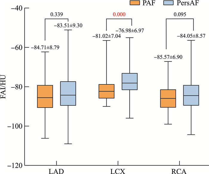
表 3 PAF组和PersAF组PCAT参数比较
Table 3. PCAT parameter comparison
X±S 项目 PAF组(95例) PersAF组(160例) P LAD-体积/mm3 1 071.70±578.36 1 146.76±650.93 0.347 LAD-FAI/HU -84.71±8.79 -83.51±9.30 0.339 LCX-体积/mm3 655.30±405.58 595.62±485.53 0.264 LCX-FAI/HU -81.02±7.04 -76.98±6.97 0.000 RCA-体积/mm3 1 485.19±943.28 1 481.60±894.00 0.911 RCA-FAI/HU -85.57±6.90 -84.05±8.57 0.095
下载: 导出CSV
-
[1] Kornej J, Börschel CS, Benjamin EJ, et al. Epidemiology of atrial fibrillation in the 21st century: novel methods and new insights[J]. Circ Res, 2020, 127(1): 4-20. doi: 10.1161/CIRCRESAHA.120.316340
[2] van der Bijl P, Kuneman JH, Bax JJ. Pericoronary adipose tissue attenuation: diagnostic and prognostic implications[J]. Eur Heart J Cardiovasc Imaging, 2022, 23(12): e537-e538. doi: 10.1093/ehjci/jeac175
[3] Antoniades C, Kotanidis CP, Berman DS. State-of-the-art review article. Atherosclerosis affecting fat: What can we learn by imaging perivascular adipose tissue?[J]. J Cardiovasc Comput Tomogr, 2019, 13(5): 288-296. doi: 10.1016/j.jcct.2019.03.006
[4] Antonopoulos AS, Antonopoulos AS, Sanna F, et al. Detecting human coronary inflammation by imaging perivascular fat[J]. Sci Transl Med, 2017, 9(398): eaal2658. doi: 10.1126/scitranslmed.aal2658
[5] Antoniades C, Patel P, Antonopoulos AS. Using artificial intelligence to study atherosclerosis, predict risk and guide treatments in clinical practice[J]. Eur Heart J, 2023, 44(6): 437-439. doi: 10.1093/eurheartj/ehac751
[6] Antonopoulos AS, Sanna F, Sabharwal N, et al. Detecting human coronary inflammation by imaging perivascular fat[J]. Sci Transl Med, 2017, 9(398): eaal2658. doi: 10.1126/scitranslmed.aal2658
[7] Lin A, Nerlekar N, Yuvaraj J, et al. Pericoronary adipose tissue computed tomography attenuation distinguishes different stages of coronary artery disease: a cross-sectional study[J]. Eur Heart J Cardiovasc Imaging, 2021, 22(3): 298-306. doi: 10.1093/ehjci/jeaa224
[8] Zhang WZ, Li PL, Chen XY, et al. The association of coronary fat attenuation index quantified by automated software on coronary computed tomography angiography with adverse events in patients with less than moderate coronary artery stenosis[J]. Diagnostics(Basel), 2023, 13(13): 2136.
[9] Vrints C, Andreotti F, Koskinas KC, et al. 2024 ESC Guidelines for the management of chronic coronary syndromes[J]. Eur Heart J, 2024, 45(36): 3415-3537. doi: 10.1093/eurheartj/ehae177
[10] Van Gelder IC, Rienstra M, Bunting KV, et al. 2024 ESC Guidelines for the management of atrial fibrillation developed in collaboration with the European Association for Cardio-Thoracic Surgery(EACTS)[J]. Eur Heart J, 2024, 45(36): 3314-3414. doi: 10.1093/eurheartj/ehae176
[11] 沈旨艳, 夏坤, 幸志洋, 等. 冠周脂肪衰减指数评估冠心病研究进展[J]. 中国医学影像技术, 2023, 39(4): 614-617.
[12] Jeudy J, Patel P, George N, et al. Assessment of coronary inflammation in antiretroviral treated people with HIV infection and active HIV/hepatitis C virus co-infection[J]. AIDS, 2022, 36(3): 399-407. doi: 10.1097/QAD.0000000000003125
[13] Visseren FLJ, Mach F, Smulders YM, et al. 2021 ESC Guidelines on cardiovascular disease prevention in clinical practice[J]. Eur Heart J, 2021, 42(34): 3227-3337. doi: 10.1093/eurheartj/ehab484
[14] Ernault AC, Meijborg VMF, Coronel R. Modulation of cardiac arrhythmogenesis by epicardial adipose tissue: JACC state-of-the-art review[J]. J Am Coll Cardiol, 2021, 78(17): 1730-1745. doi: 10.1016/j.jacc.2021.08.037
[15] Ma GJ, Guo FQ, Hu J, et al. Association of pericoronary adipose tissue with atrial fibrillation recurrence after ablation based on computed tomographic angiography[J]. Jpn J Radiol, 2023, 41(9): 955-964. doi: 10.1007/s11604-023-01426-x
[16] Kim HW, Belin de Chantemèle EJ, Weintraub NL. Perivascular adipocytes in vascular disease[J]. Arterioscler Thromb Vasc Biol, 2019, 39(11): 2220-2227. doi: 10.1161/ATVBAHA.119.312304
[17] 李晓乐, 谢丽响, 严卉, 等. 冠周脂肪及甘油三酯-葡萄糖指数对不同类型心房颤动射频消融术后复发的预测价值[J]. 临床心血管病杂志, 2024, 40(8): 669-674. doi: 10.13201/j.issn.1001-1439.2024.08.013
[18] Nogami K, Sugiyama T, Kanaji Y, et al. Association between pericoronary adipose tissue attenuation and outcome after second-generation cryoballoon ablation for atrial fibrillation[J]. Br J Radiol, 2021, 94(1128): 20210361. doi: 10.1259/bjr.20210361
[19] Mekhael M, Marrouche N, El Hajjar AH, et al. The relationship between atrial fibrillation and coronary artery disease: Understanding common denominators[J]. Trends Cardiovasc Med, 2024, 34(2): 91-98. doi: 10.1016/j.tcm.2022.09.006
-
计量
- 文章访问数: 24
- 施引文献: 0