
Analysis of reasons for missed diagnosis in ultrasound diagnosis of inferior vena cava anomalous connection
-
摘要: 目的 探讨经胸超声心动图(TTE)诊断下腔静脉异位连接的漏诊原因。 方法 回顾性分析2015年3月—2024年1月于西京医院超声医学科行TTE检查患者的临床资料,共发现31例下腔静脉异位连接,全部经CT血管成像(CT angiography,CTA)、心血管造影和(或)外科手术证实,分析其超声心动图特征、诊断的准确率及漏诊率。 结果 下腔静脉异位连接共31例,其中右位下腔静脉18例,左位下腔静脉11例,双下腔静脉2例。19例(61%,19/31)经奇静脉回流入右位上腔静脉、6例(19%,6/31)经半奇静脉-奇静脉回流入右位上腔静脉、5例(16%,5/31)经半奇静脉回流入左位上腔静脉、1例(3%,1/31)直接回流入冠状静脉窦。超声确诊22例(71%,22/31)、漏诊9例(29%,9/31)。29例(94%,29/31)合并心内畸形,2例(6%,2/31)为孤立性下腔静脉异位连接,心内结构正常。合并畸形中房间隔缺损(ASD)9例、单心房(SA)7例、单心室(SV)6例、肺动脉狭窄(PS)6例、室间隔缺损(VSD)5例、永存左上腔静脉(LSVC)5例、动脉导管未闭(PDA)4例、肺静脉异位引流(APVC)3例、房室间隔缺损(AVSD)2例、右室双出口(DORV)1例、无顶冠状静脉窦综合征(UCSS)1例、肺动脉闭锁(PA)1例、二叶式肺动脉瓣(BPV)1例、右上腔静脉缺如1例。 结论 TTE容易漏诊下腔静脉异位连接。全面扫查剑突下下腔静脉长轴及短轴切面观察下腔静脉回流入右房情况,胸骨旁胸主动脉长轴及短轴切面观察胸主动脉旁有无代偿扩张的奇静脉或半奇静脉,胸骨旁、剑突下、胸骨上窝、锁骨上窝各个上腔静脉切面观察上腔静脉内径是否增宽,血流速度是否加快、有无代偿扩张的奇静脉或半奇静脉汇入,联合CTA或心血管造影可降低漏诊率。Abstract: Objective To explore the reasons for missed diagnosis of inferior vena cava anomalous connection using transthoracic echocardiography(TTE). Methods Retrospective analysis of patients who underwent TTE in the Ultrasound Medicine Department of Xijing Hospital from March 2015 to January 2024. A total of 31 cases of inferior vena cava anomalous connections were discovered, all of which were confirmed by CT angiography(CTA), Cardiac angiogram, and/or surgical procedures. The echocardiographic characteristics, diagnostic accuracy, and missed diagnosis rate were analyzed. Results Among the 31 cases of inferior vena cava ectopia, there were 18 cases of right inferior vena cava, 11 cases of left inferior vena cava, and 2 cases of bilateral inferior vena cava. The 19 cases(61%, 19/31) returned to the right superior vena cava through the azygos vein, 6 cases(19%, 6/31) returned to the right superior vena cava through the hemiazygos vein-azygos vein, 5 cases(16%, 5/31) returned to the left superior vena cava through the hemiazygos vein, and 1 case(3%, 1/31) directly returned to the coronary sinus. Ultrasound confirmed 22 cases(71%, 22/31) and missed 9 cases(29%, 9/31). 29 cases(94%, 29/31) had concomitant intracardiac malformations, and 2 cases(6%, 2/31) had isolated inferior vena cava ectopic connections with normal internal structures. There were 9 cases of atrial septal defect(ASD), 7 cases of single atrium(SA), 6 cases of single ventricle(SV), 6 cases of pulmonary artery stenosis(PS), 5 cases of ventricular septal defect(VSD), 5 cases of persistent left superior vena cava(LSVC), 4 cases of patent ductus arteriosus(PDA), 3 cases of anomalous pulmonary venous drainage(APVC), 2 cases of atrioventricular septal defect(AVSD), 1 case of right ventricular double outlet(DORV), 1 case of unrounded coronary sinus syndrome(UCSS), 1 case of pulmonary artery occlusion(PA), 1 case of bicuspid pulmonary valve(BPV), and 1 case of absence of right superior vena cava. Conclusion TTE is more likely to miss the diagnosis of inferior vena cava anomalous connection. A comprehensive scan of the long and short axis sections of the inferior vena cava under the xiphoid process was conducted to observe the reflux of the inferior vena cava into the right atrium. The long and short axis sections of the thoracic aorta near the sternum were used to observe the presence of compensatory dilation of the azygos or Hemiazygos veins. The sections of the superior vena cava near the sternum, under the xiphoid process, suprasternal fossa, and supraclavicular fossa were used to observe whether the inner diameter of the superior vena cava had widened, whether the blood flow velocity had increased, and whether there was compensatory dilation of the azygos or Hemiazygos veins merging. Combined with CTA or Cardiac angiogram, the missed diagnosis rate could be reduced.
-

-
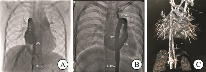
图 1 下腔静脉异位连接的心血管造影及CTA表现
Figure 1. Angiography and CTA findings of ectopic connection of inferior vena cava
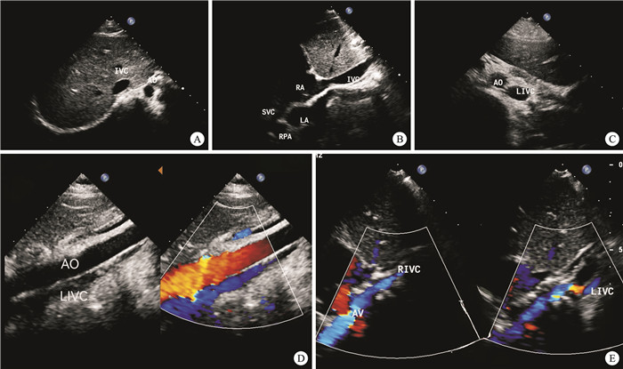
图 2 正常下腔静脉与下腔静脉异位连接剑突下短轴和长轴的超声表现
Figure 2. Ultrasonographic manifestations of the minor axis and major axis of the xiphoid process
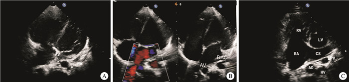
图 3 与下腔静脉异位连接时奇静脉、半奇静脉的超声表现
Figure 3. Ultrasonic manifestations of azygos vein and semi-azygos vein when heterotopic connection with inferior vena cava is made
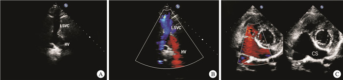
图 4 下腔静脉异位连接时左位下腔静脉经半奇静脉延续连接汇入左位上腔静脉的超声表现
Figure 4. Ultrasonic manifestations of the left inferior vena cava merging into the left superior vena cava
表 1 9例下腔静脉异位连接的超声诊断漏诊结果
Table 1. Nine cases of missed diagnosis results of ultrasound diagnosis of ectopic connection of inferior vena cava
病例 男/女 年龄/岁 超声诊断心内畸形 心血管造影/CTA/手术确诊 漏诊下腔静脉异位连接的异位引流途径 1 男 57 心内结构正常 心血管造影手术 R-IVC经AV延续回流入R-SVC 2 女 3 VSD 心血管造影手术 双-IVC经AV延续回流入R-SVC 3 女 4 PDA 心血管造影手术 R-IVC经AV延续回流入R-SVC 4 女 25 PDA、ASD 心血管造影手术 R-IVC经AV延续回流入R-SVC 5 女 34 PDA 心血管造影手术 L-IVC经HV延续回流入L-SVC 6 男 1 APVC、ASD CTA、手术 L-IVC经HV-AV延续回流入R-SVC 7 女 33 APVC、ASD CTA、手术 R-IVC经AV延续回流入R-SVC 8 男 5 VSD、ASD 手术 R-IVC经AV延续回流入R-SVC 9 男 24 SV、SA、PS CTA R-IVC经AV延续回流入R-SVC 合计 男4例
女5例8例合并心内畸形 全部经CTA/心血管
造影/手术确诊7例经AV延续回流入R-SVC
1例经HV延续回流入L-SVC
1例经HV-AV延续回流入R-SVC注:VSD室间隔缺损;PDA动脉导管未闭;ASD房间隔缺损;APVC肺静脉异位引流;SV单心室;SA单心房;PS肺动脉瓣狭窄。  下载: 导出CSV
表 2 31例下腔静脉异位连接的异位引流途径
Table 2. Ectopic drainage pathway of ectopic connection of inferior vena cava in 31 cases
异位引流途径 总计(31例) 占比 经AV延续回流入R-SVC 19 61% 经HV-AV延续回流入R-SVC 6 19% 经HV延续回流入L-SVC 5 16% 直接回流入CS 1 3%
下载: 导出CSV
-
[1] Malaki M, Willis AP, Jones RG. Congenital anomalies of the inferior vena Cava[J]. Clin Radiol, 2012, 67(2): 165-171. doi: 10.1016/j.crad.2011.08.006
[2] Kandpal H, Sharma R, Gamangatti S, et al. Imaging the inferior vena cava: a road less traveled[J]. Radiographics, 2008, 28(3): 669. doi: 10.1148/rg.283075101
[3] Eldefrawy A, Arianayagam M, Kanagarajah P, et al. Anomalies of the inferior vena Cava and renal veins and implications for renal surgery[J]. Cent Eur J Urol, 2011, 64(1): 4-8.
[4] Minniti S, Visentini S, Procacci C. Congenital anomalies of the venae cavae: embryological origin, imaging features and report of three new variants[J]. Eur Radiol, 2002, 12(8): 2040-2055. doi: 10.1007/s00330-001-1241-x
[5] 闫庆. 下腔静脉离断的研究进展[J]. 临床医药实践, 2024, 33(7): 538-540.
[6] 颜华英, 何丽红, 张春国, 等. 产前超声诊断胎儿下腔静脉异常连接[J]. 中国医学影像学杂志, 2023, 31(7): 756-760. doi: 10.3969/j.issn.1005-5185.2023.07.016
[7] Cizginer S, Tatli S, Girshman J, et al. Thrombosed interrupted inferior vena Cava and retroaortic left renal vein mimicking retroperitoneal neoplasm[J]. Abdom Imaging, 2007, 32(3): 403-406. doi: 10.1007/s00261-006-9052-9
[8] Truty MJ, Bower TC. Congenital anomalies of the inferior vena Cava and left renal vein: implications during open abdominal aortic aneurysm reconstruction[J]. Ann Vasc Surg, 2007, 21(2): 186-197. doi: 10.1016/j.avsg.2006.10.014
[9] Bass JE, Redwine MD, Kramer LA, et al. Spectrum of congenital anomalies of the inferior vena Cava: cross-sectional imaging findings[J]. Radiographics, 2000, 20(3): 639-652. doi: 10.1148/radiographics.20.3.g00ma09639
[10] Tacy TA, Silverman NH. Systemic venous abnormalities: embryologic and echocardiographic considerations[J]. Echocardiography, 2001, 18(5): 401-413. doi: 10.1046/j.1540-8175.2001.00401.x
[11] 赫瑞, 朱铭. 先天性下腔静脉畸形影像学诊断[J]. 中国临床医学影像杂志, 2009, 20(9): 708-711. doi: 10.3969/j.issn.1008-1062.2009.09.014
[12] De Rosa V, Catalano O, de Lutio di Castelguidone E, et al. Segmental septation of the inferior vena Cava[J]. Am J Roentgenol, 2005, 185(5): 1377. doi: 10.2214/AJR.04.1827
[13] Baldridge ED Jr, Canos AJ. Venous anomalies encountered in aortoiliac surgery[J]. Arch Surg, 1987, 122(10): 1184-1188. doi: 10.1001/archsurg.1987.01400220094018
[14] Lim JSL, McCrindle BW, Smallhorn JF, et al. Clinical features, management, and outcome of children with fetal and postnatal diagnoses of isomerism syndromes[J]. Circulation, 2005, 112(16): 2454-2461. doi: 10.1161/CIRCULATIONAHA.105.552364
[15] 陈东, 冯俊, 江永进, 等. 伴下腔静脉滤器经上腔静脉路径成功消融右侧旁路1例[J]. 临床心血管病杂志, 2024, 40(1): 78-80. doi: 10.13201/j.issn.1001-1439.2024.01.015
[16] 段红永, 汪忠镐. 下腔静脉畸形的胚胎发育及临床意义[J]. 国际外科学杂志, 2008, 35(12): 854-856. doi: 10.3760/cma.j.issn.1673-4203.2008.12.023
[17] Sheth S, Fishman EK. Imaging of the inferior vena Cava with MDCT[J]. AJR Am J Roentgenol, 2007, 189(5): 1243-1251. doi: 10.2214/AJR.07.2133
[18] 罗晓莉, 江丽, 朱建平. 彩色多普勒超声诊断先天性下腔静脉畸形的临床应用价值[J]. 中华医学超声杂志(电子版), 2014, 11(2): 125-130.
-
计量
- 文章访问数: 19
- 施引文献: 0